Showing posts with label Board Question. Show all posts

Thursday, October 7, 2010

Board question: Transplant answer


The correct answer is D.
The criteria for expanded-criteria donors (ECD) are:
  • age greater than 60 years old
OR
  • age 50 to 59 years old and at least two of the following:
  1. History of hypertension
  2. Terminal creatinine > 1.5mg/dL
  3. Death from a cerebral vascular accident
This new category of renal transplantation donors was introduced in 2002 to help expand the pool of potential kidney donors. ECD are associated with a 70% higher risk of graft failure compared to non-ECD transplants. About 20% of deceased-donor transplants in 2008 are ECD kidneys in the US. The 3-year graft survival for ECD transplants was 67% in 2003. By comparison, the 3-year graft survival for non-ECD and living-donor transplants is 80 and 88%, respectively. Since the supply of suitable kidney transplants fails to match the increasing demands, nephrologists will need to determine if ECD transplants are appropriate for their patients. The decision must be individualized and patient-centered, since we have not identified the cohort for whom ECD transplants will most benefit. Expect to see a question regarding ECD transplants on the renal boards!

Michael Lattanzio DO

Friday, August 27, 2010

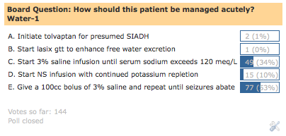
Board question: Water-1 answer


This was an actual case I saw and thought it generated a multitude of interesting dilemmas. When I initially examined the patient she was in status epilepticus. This, by itself, mandated the immediate correction of her serum sodium. But what was the best way to achieve the correction without adversely affected the patient? This was my thought process…

The patient is 100kg women, so her TBW should be 50 Liters.
(using the Watson Formula)

I decided to separate her water and solute gains/losses to determine what her total body sodium was at that instance.


She had already received 2L of normal saline but lost 3L in urine, so net water loss of 1L. 1L of free water loss would cause that serum sodium to rise 2.5meq/L.

Calculation: 120 (desired Na)-115 (actual Na)/120 (desired Na) times 50L (TBW) is equal to roughly 2L. Therefore, 2L of free water loss would raise serum sodium 5 meq/L.

Therefore, for each 1L of water loss, the serum sodium would rise 2.5 meq/L. (5meq/L divided by 2)

So, just based on water loss, her serum sodium has risen 2.5 meq/L already, so the serum sodium is 117.5 meq/L.
Next, I calculated the impact of solute gain. Total solute for a 100kg woman with a serum sodium of 115 would be 5750 (50L times 115meq/L). I estimated that she gained 133 meq of solute… INPUT: 2L of normal saline 388meq KCL 80meq Total input 468 meq solute input LOSS: Urine sodium of 75 meq/L times 3L urine output is 225 meq Urine potassium of 10 meq/L times 3L urine output is 30 meq Total loss: 255meq

So, total solute input (388)-total solute output (255) equals a gain of 133meq of solute.

How does this impact the serum sodium?
Total body solute 5750 plus gain of 133 equals 5865 5865 meq (New Total Body Solute) divided by TBW (50 L) equals 117.3 Therefore, serum sodium rose 2.3 based on solute gain alone.

If you add the affect of free water loss and solute gain, I anticipated that the serum sodium at that instance was 120 meq/L (2.5 meq/L from water loss and 2.3 from solute gain plus starting sodium of 115). So, I thought that the serum sodium was already in the “safe” zone and that the seizures were related to alcohol withdrawl.

I decided NOT to use 3% saline, and continue with gentle potassium repletion at that time (depite the intensivist’s insistence). What this physician failed to realize was that KCl has the same osmotic potential as sodium!

In fact, there are case reports of central pontine myelinosis induced by rapid correction of hyponatremia via potassium repletion alone.


The patient’s repeat sodium was 121! Her serum sodium stabilized over the coarse of a few days with mere potassium repletion. Her seizures persisted and patient eventually required anti-epileptic therapy.

Given the complexities of this case, there is not a perfect strategy for management. The purpose was to generate thoughts and discussions on the intricacies of hyponatremia management. Would you have managed her differently? Let me know your thoughts…

Michael Lattanzio, DO

Tuesday, August 24, 2010

Board question of the week: Water-1

A 40-year-old woman is rushed to ER for change in mental status. She has a history of multiple sclerosis and alcohol abuse. On arrival to ER, she exhibits 3 stereotyped, tonic-clonic seizure that resolve with ativan. The initial metabolic panel is listed below:
Na- 115, K- 1.6, Cl- 56, HCO3- 40, BUN- 10, Cr- 0.8, Serum osmolality- 240 Her husband arrives at the ER and reports heavy alcohol use over the last few weeks. He brings her medication list, which includes: Protonix, Tysabri (natalizumab, anti-alpha-4-integrin antibody), and Prednisone. Blood work from 3 months prior show a normal sodium level. She received 2L of normal saline in the ER, 80 meq of KCl, and one dose of lasix. Her urine output has been 3L so far. Urine electrolytes are obtained (Una 75, Uk 10). She weighs 100kg and on examination the patient is noted to be in “status”. Nephrology is consulted for the management of her hyponatremia.

How should this patient be managed acutely?
Possible answers are listed in the poll on the upper right hand column.

The answer and explanation will be posted on Friday August 27th

Michael Lattanzio DO

*RFN board questions are meant to help introduce concepts about nephrology related diseases and do not represent actual questions seen on the ABIM exam.

Tuesday, August 3, 2010

Board Review; Pregnancy answer

Each of the following choices is expected to occur during pregnancy except for A and D. Let’s go through each of the possible choices.

A. This in partially incorrect. Hyperventilation (with resultant hypocapnea, not hypercapnea) occurs during pregnancy and is largely a consequence of stimulation of the respiratory center via progesterone. Therefore, in pregnancy you typically see a chronic respiratory alkalosis which is compensated by renal excretion of bicarbonate. Luckily, in pregnancy the oxygen-dissociation curve is not dramatically altered due to increased 2,3 DPG levels in pregnancy and normalization of blood pH through renal compensation.

B. This is correct. During pregnancy the intestinal absorption of calcium nearly doubles as early as week 12 of pregnancy and is likely the major maternal adaptation to meet the fetal need for calcium. As intestinal calcium absorption increases, extra calcium is either deposited in the fetal skeleton, the maternal skeleton or is excreted by the kidneys. Renal calcium excretion is increased as early as the 12th week of gestation and 24 hour urine values (corrected for creatinine excretion) can exceed the normal range. Conversely, fasting urine calcium values are normal or low, confirming that the hypercalciuria is a consequence of the enhanced intestinal calcium absorption. Pregnancy is recognized as a risk factor for kidney stones and the absorptive hypercalciuria of pregnancy is one reason for this.

C. This is also correct. During pregnancy kidney size can increase by 20%. This increase in kidney size also corresponds to an increase in GFR and renal blood flow.

D. This is incorrect. Remember that filtration fraction (FF) is the ratio of glomerular filtration rate (GFR) to renal blood flow (RBF). The filtration fraction represents the proportion of fluid reaching the kidneys which eventually passes into the tubular lumen. In pregnancy, although the GFR is increased, RBF is increased to a greater extent. Therefore, FF actually decreases in pregnancy. Since the increase in GFR and RBF is not associated with an increase in FF, there is no risk of secondary FSGS in pregnancy. This is supported by small studies.

E. This is correct. During pregnancy, urinary protein excretion increases from normal nonpregnant levels of 60–90 mg/24 hours to 180–250 mg/24 hours in the third trimester. Even if slightly increased during pregnancy, urine protein excretion rarely reaches levels that are detected by usual screening methods, such as 1+ on urinary dipstick (i.e., 30 mg/dL, which is roughly equivalent to 300 mg in 24 hours). Although the mechanism for this possible increase has not been established, it seems likely that absorption of filtered protein in the proximal tubule is reduced.

F. This is correct as well. During pregnancy, physiologic changes occur in volume- and osmoregulation that effect plasma osmolality and sodium concentration. During a normal pregnancy, the average plasma osmolality decreases by 5-10 mmol, and the sodium concentration is decreased by 5 mmol/l. This drop in plasma osmolality has been explained by a phenomenon termed ‘reset osmostat’. A ‘reset osmostat’ is when the osmotic threshold above which ADH-release and a thirst stimulus occur changes. This threshold is decreased to a lower steady state value during normal pregnancy. The mechanism that causes the reset-osmostat phenomenon is unknown.

On a lighter note, the “Gross Clinic” has completed its restoration and is currently on exhibit at the Philadelphia Museum of Art. It is a must-see for any devotee of medicine.

Michael Lattanzio DO

Friday, July 16, 2010

Board question: PD-1 answer

The best answer is E. Encapsulating sclerosing peritonitis (ESP)

This patient presented with true UF failure (<400cc UF volume on a 4hr PET using 4.25% with 2L dwell volume- "rule of 4's").

The differential diagnosis of true UF failure can be separated by PET results:

Decreased D/P Cr
  1. Encapsulating Sclerosing Peritonitis (ESP)
  2. Adhesions
Stable D/P Cr
  1. Increased lymphatic absorption
  2. Dialysate leak
  3. Catheter Malposition
  4. Decreased Transcellular Transport
Increased D/P Cr
  1. Peritonitis
  2. High transport status
ESP is a progressive, intra-abdominal, inflammatory process that results in layers of fibrous tissue that encapsulate the viscera, thereby compromising the motility and function of the bowel. The length of time on PD is the most important risk factor for ESP, ie, the more time on PD, the greater likelihood of developing ESP. Also, high transporters may be at heightened risk for ESP compared to low or average transporters.
The presentation of ESP may include:
The CT abdomen/pelvis shows dense sheets of fibrous material in the peritoneum.
The diagnosis of ESP mandates the immediate cessation of PD. Surgery is very challenging and is usually not recommended. Steroids and tamoxifen have shown some efficacy in case studies, but this has not been systematically studied. Nutritional support with TPN is often necessary.

Michael Lattanzio DO

*RFN board questions are meant to help introduce concepts about nephrology related diseases and do not represent actual questions seen on the ABIM exam.

Monday, July 12, 2010

Board question of the week: PD-1

A 55-year-old woman with ESRD secondary to lupus nephritis presents to the PD clinic with volume overload. She reports decreased UF volume, despite the use of 4.25% dextrose exchanges over the last few weeks. She has been on PD for the last 12 years. Historically, she has been able to maintain her weight, despite being a rapid transporter. She denies any abdominal pain or cloudy fluid. An abdominal film shows proper placement of the PD catheter in the lower pelvis. A catheter assessment shows no flow obstruction. A 4 hour PET using 4.25% dextrose solution results in the ultrafiltration of 350cc on a 2L dwell. The D/P Cr level is reduced from her previous studies.

What is the most likely explanation for these results? Possible answers are listed in the poll on the upper right hand column.

The answer and explanation will be posted on Friday July 16th

Michael Lattanzio DO

*RFN board questions are meant to help introduce concepts about nephrololgy related diseases and do not represent actual questions seen on the ABIM exam.

Monday, June 21, 2010

Board question: Transplant-1 answer


The vast majority of people answered correctly; the best answer is D.


Approximately 3-5% of patients with Alport's syndrome develop de novo anti-glomerular basement membrane (GBM) disease in the transplanted kidney. Alport’s syndrome is a genetic disorder that results from mutations in the genes encoding the alpha-3, alpha-4, or alpha-5 chains of type IV collagen. Following transplantation, recipients can become alloimmunized and develop antibodies to the normal chains of type IV collagen in the basement membrane of the donor kidney (hence, the linear pattern of staining noted on IF). Treatment is not standardized, but generally consists of plasmapharesis, +/- cyclophosphamide and steroids. Long-term allograft survival is poor and re-transplantation carries a high risk of anti-GBM recurrence. For the boards, remember the post-transplant association between Alport’s syndrome and de novo anti-GBM disease.

Link to NEJM review which contains above picture


Michael Lattanzio DO

Thursday, June 17, 2010

Board question of the week: Transplant-1

An 18-year-old white man presents to the renal transplant clinic with complaints of fatigue and nausea for the last week. He was the recipient of a deceased donor kidney transplant 12 months prior and has experienced stable allograft function (Cr 1.2 mg/dL) thereafter. His original renal disease was Alport’s syndrome. His current immunosuppressive regimen consists of tacrolimus and mycophenolate mofetil. He reports strict compliance with this regimen. The nephrologist orders bloodwork:

Bun-50 Cr-5.0 UA-3+ blood 3+ protein dysmorphic RBCs noted
Spot protein/creatinine ratio- 5g/d
C3/C4- normal, FK506 level- 10

Renal transplant ultrasound- no gross abnormality

ANCA pending

Given the worsening renal function, a transplant biopsy is immediately performed. The light microscopy and immunofluorescence appear below:


The answer and explanation will be posted on Monday June 21st

Michael Lattanzio DO


*RFN board questions are meant to help introduce concepts about nephrology related diseases and do not represent actual questions seen on the ABIM exam.

Monday, June 7, 2010

Board question: Hypertension-1 answer


The best answer is D.


Lifton et al. described a single gene mutation on the hormone-binding domain of the mineralocorticoid receptor (MCR). Individuals with this missense mutation develop early-onset hypertension with characteristic low renin and aldosterone levels. The mutation causes the MCR to be constitutively active, regardless of aldosterone levels, and transforms the MCR so that steroid hormones that are typically antagonistic become agonistic (in particular, progesterone and cortisone).

During pregnancy, when progesterone levels increase 100-fold, the MCR becomes hyperactive and leads to sodium reabsorption and potassium secretion via the principal cell of the distal collecting tubule (Figure above). The avid sodium retention and volume expansion appropriately suppress renin and aldosterone levels. This condition is not associated with proteinuria, edema, or neurologic changes, which distinguishes it from pre-eclampsia.

Given the low levels of aldosterone, this condition is refractory to standard medical therapy aimed at reducing aldosterone levels through RAAS blockade. In fact, mineralocorticoid receptor antagonists can actually exacerbate hypertension in this condition! Delivery of the fetus may be necessary to treat severe, refractory hypertension during pregnancy.

Essential hypertension results from a complex interplay of both genetic and environmental influences. Uncommonly, an isolated genetic mutation can engender hypertension, like in this case. These monogenic forms of hypertension affect either electrolyte transport in the distal nephron, or the synthesis and/or activity of mineralocorticoids, leading to a common final pathway of increased distal tubular reabsorption of sodium and chloride, volume expansion, and hypertension.

Other low renin, monogenic forms of hypertension include:
  • Familial Hyperaldosteronism Type 1 (aka, glucocorticoid-remediable aldosteronism)
  • Familial Hyperaldosteronism Type II
  • Syndrome of apparent mineralocorticoid excess
  • Liddle syndrome
  • Pseudohypoaldosteronism type II (aka, Gordon syndrome)
  • Congenital Adrenal Hyperplasia

They provide excellent fodder for any number of board-style questions!

Michael Lattanzio DO

Thursday, June 3, 2010

Board question of the week: Hypertension-1

A 26-year-old primigravid woman is admitted during her 34th week of pregnancy with severe, refractory hypertension. She was noted to have mild hypertension prior to pregnancy. Her family history is remarkable for pregnancy-related hypertension in her aunt. Her blood pressure decreased early in the course of the pregnancy, but then rose dramatically, reaching 170/100 mmHg at 29 weeks despite antihypertensive therapy. She denies any edema or neurologic changes.

Her chemistries are listed below:

Na-143 K-3.6 Cl-110 HCO3-28 BUN-8 Creat-0.5 Uric Acid-3

Urinalysis negative for blood and protein

Urine electrolytes:

Urine sodium 20 meq/L, Urine potassium 75meq/L

Renin and Aldosterone level were both suppressed

Due to worsening blood pressure, a Caesarian section was performed, and the patient delivered a healthy baby girl. Her blood pressure control stabilized shortly after delivery.


The answer and explanation will be posted on Monday July 7th

Michael Lattanzio DO

*RFN board questions are meant to help introduce concepts about nephrology related diseases and do not represent actual questions seen on the ABIM exam.