Showing posts with label Finnian McCausland. Show all posts

Sunday, January 22, 2012

Azathioprine in pregnancy

We recently discussed this issue in conference and I thought it might be worth sharing a few interesting points:

Azathioprine is normally converted to the active metabolite 6-mercaptopurine. However, in pregnancy the placenta can metabolize azathioprine to thiouric acid, an inactive metabolite. In addition the fetal liver does not have inosinatopyrophosphorylase which therefore largely protects the fetus from exposure to active compounds.

Azathioprine has been classed as FDA category D - positive evidence of risk.
Despite conflicting data, general expert opinion suggests that azathioprine may be considered for use during pregnancy in certain situations (where the potential benefits outweigh potential risks).

In terms of breast feeding, in a small study, active metabolites have been detected in small quantities in breast milk. Overall the significance of this remains to be fully determined. Manufacturers have taken the official stance to warn against breast feeding while taking the drug.


As with all considerations of alterations in immunosuppressive dosing, careful consideration of risks and benefits should be explored in detail with the treating physician before any change is undertaken.

Sunday, December 18, 2011

The Origins of Renal Physiology, a National Course for Renal Fellows

This fall I attended the fourth annual Origins of Renal Physiology course for nephrology fellows at the Mount Desert Island Biological Laboratory (MDIBL). The course was designed to allow fellows an opportunity to connect with the historical roots of renal physiology and to gain perspective through comparative physiology. The founder of this blog, Nathan Hellman, attended the first course and blogged about his experience in 2008.

The course took place on the coast of Maine at the MDIBL, a place with great historical and modern day significance for renal physiology. George Dorr, who founded Acadia National Park with John D. Rockefeller, had the foresight to recognize the potential value of the island for the scientific community. He offered land for two labs, the MDIBL and the Jackson Laboratory (which also continues to operate on the island with tremendous success). The MDIBL is where E. K. Marshall discovered and described active tubular transport in the goosefish in the 1920’s. It is where Homer Smith wrestled with the concepts of clearance, GFR, and effective renal blood flow and worked out calculations based on his studies in marine animals. He did ground breaking experiments in the lab and wrote a number of manuscripts and books in his cottage on the campus describing these fundamental concepts. For 35 years, he spent his summers working at the MDIBL. Roberts Pitts called this time the “Smithian Era of renal physiology”. Homer Smith brought many others to the island to work on renal physiology, and over the years a number of important discoveries have been made at the MDIBL. The lab continues to be a place of excellence, active in research.

The annual course brings fellows back to this historical place and places them under the mentorship of a distinguished faculty. Mark Zeidel serves as the course director and every year he brings in a remarkable faculty from across the country to distill fundamental concepts of renal physiology in a historical and comparative context. They do this by dividing the course up into six modules. Each fellow participates in three of the six modules with each module lasting for two days. The modules split the kidney up into functional units beginning with the glomerulus and continuing on through the proximal tubule, the loop of Henle, and the distal tubule. They also covered salt and water homeostasis and renal genetics. On the first day of each module, we worked with the faculty in the lab performing experiments using classic physiology model systems such as the toad bladder, the shark rectal gland, the zebrafish, and the xenopus oocyte. We developed a presentation based on our experiments to share with the group on the morning of the second day of each module. Even though we only work with three of the modules each year, we learned what the other groups were doing through the peer presentations. The afternoons after the presentations were spent exploring the island and Acadia National Park. There were organized outings for hiking, biking, and kayaking. We finished the week with a big lobster bake at a park on the seawall.

I have had the great pleasure of attending the course each of the last two years, participating in all six modules. I told my wife when I got home each time that I felt like a kid at camp. Only instead of the usual camp games, we were playing with kidneys and urine. These were tremendous experiences that I will carry with me throughout my career in nephrology. It was great to meet and work with faculty and fellows from across the country. I came away with an enhanced understanding of physiology and a renewed enthusiasm for all things renal. As you make your schedules out for next year, I would encourage you to consider setting aside some time to attend next year’s course, September 2-9.

Posted by Michael Hovater MD

Sunday, December 4, 2011

Renal disease and HIV

Perhaps the most renowned renal disease associated with HIV infection is the collapsing variant of FSGS. This typically presents with the nephrotic syndrome and has been associated with relatively advanced HIV infection, with low CD4 counts and higher viral loads. HIV infection of tubular cells may play a direct role in the pathogenesis, with some supporting evidence from animals studies in which transgenic mice were created with HIV constructs in their genome – they developed proteinuria and similar histological findings of collapsing glomerular tufts and interstitial disease. Hypertension appears to be less common, possibly due to an association with salt wasting.

In addition there are many other renal diseases that can be associated with HIV infection, its treatment or associated conditions. An interesting review can be found here.

Below are some of the differentials on that long list:

Many of the anti-retrovirals have been associated with nephrotoxicity in various forms. Further information can be found on previous posts.

Similarly, various antibiotics and other anti-microbials used to treat or prophylaxis this population must be kept in mind (e.g. Bactrim, amphotericin, pentamidine etc.). Another potential problem over the longer term is the diabetogenic potential from various protease inhibitors.

Co-infection with other diseases, particularly HBV and HCV should be investigated. This opens a whole other list of differentials in terms of renal disease – e.g. membranous associated with HBV and syphilis; cryoglogulin and membranoproliferative disease associated with HCV. An immune complex mediated disease with predominance of IgA has also been described, as have various forms of lupus like clinical cases.

One other rare consideration is thrombotic thrombocytopaenic purpura, which may result from HIV induced endothelial injury.

Just some of the differentials to keep in mind when asked to see a patient with kidney disease and HIV infection.

Monday, October 24, 2011

Bactrim prophylaxis with high-dose steroid use?


This is a question we face quite often in the Nephrology clinic when we are commencing patients on high dose steroids for glomerular and other systemic inflammatory diseases.

I came across a couple of interesting retrospective studies while trying to find some evidence to help guide the decision-making process.

The first study analyzed 116 cases of pneumocystis pneumonia in non-HIV infected patients. The major common exposure was steroid treatment in the month preceding presentation (90.5% of patients) – the median dose was equivalent to 30mg prednisone for a median of 12 weeks before presentation.


The second reported risk factors in 41 cases in a French centre. Mortality was 29%; 85% had received steroids at a median daily steroid dose equivalent to 15mg prednisone. Only 17% had received prophylaxis.


The third used a matched case-control type analysis in patients with SLE – they reported 15 cases of pneumocystis infection; risk factors for infection compared to 60 matched controls included higher disease activity, renal involvement and higher mean cumulative steroid dose (49 vs 20mg/day; p<0.01).

Finally a Cochrane review of RCTs of prophylaxis against PCP (versus none/placebo/non-PCP agents) suggested that prophylaxis was warranted when the risk of PCP infection is higher than 3.5% for adults – the authors suggested this included the following groups – “recipients of solid organ or allogeneic bone allografts for the first 6 months after transplant and, for the latter, throughout the period of immunosuppression, as well as in patients with acute lymphoblastic leukemia and Wegener granulomatosis”


There are a number of factors to be considered when trying to answer this difficult scenario – the dose of steroids used, the duration of treatment, use of other immunosuppressive drugs (i.e. the net state of immunosuppression), medical co-morbid conditions and infection history.
Many experts suggest prophylaxis for patients with rheumatological diseases requiring treatment with ≥20mg prednisone for greater than one month when used in combination with another immunosuppressive drug. Overall the pros and cons of PCP prophylaxis must ultimately be judged on a case-by-case basis.

Thursday, October 13, 2011

MDIBL - ASN reunion


Date for your diary

Dr Zeidel and the MDIBL crew wish to invite all previous attendees to the Second Annual Reunion of the Mount Desert Island Biological Laboratory National Course for Renal Fellows.

This is a great opportunity for anyone attending Kidney Week to catch up and network with former students and faculty of this wonderful course. More information in the course itself can be found here.

The details of the reunion are below:

Friday, November 11, 2011
6:45 p.m. – 7:45 p.m.
Philadelphia Marriott Downtown
Rooms 309/310


This reunion is sponsored by the American Society of Nephrology.

Sunday, October 2, 2011

Renal allograft rupture

Renal allograft rupture has been defined as a split in the renal capsule, associated with haemorrhage, when no identifiable injury was noted at the time of harvesting.

Renal allograft rupture is a rare, but life-threatening, complication of renal allograft placement. It most often occurs in the first two weeks post-transplant and usually requires immediate surgical intervention. The classical presentation is with sudden onset of pain at the graft site, shock and oliguria.


The pathophysiology appears to be secondary to increased intra-renal pressure and and associated secondary injury. The increased pressure can result in splitting of the capsule, usually along the longitudinal outer border, with resultant haemorrhage.

The main putative aetiologies have been described in many case series including this one, and include acute rejection, acute tubular necrosis, renal vein trombosis, increasing PRA, increasing age, biopsy of the graft and infection.

In my quick analysis of published cases series over the last ten years, the crude incidence of RAR appears to be around 1.11%. Of these cases, the majority were caused by acute rejection (55.8%), ATN (34.7%) and renal vein thrombosis (7.4%).

Immediate management involves supportive care and resuscitation, followed by containment of the ruptured area by surgical means or sometimes graft removal. The literature supports an atempt at graft salvage, with many successful outcomes reported to date. Thankfully it remains a relatively rare diagnosis, but definitely one we should all be aware of.

Friday, September 16, 2011

Sodium and seizures?

The development of acute hyponatraemia can have profound neurological consequences. After creation of an osmotic gradient between the intravascular compartment and the intracellular compartment, water must somehow gain access to the brain tissue.

This appears to be mediated, at least partially, by aquaporin 4, based on evidence from mouse knockout models. Those mice deficient in AQP4 had significantly less brain oedema compared to wild-type animals after the induction of acute hyponatraemia.

In very acute situations the brain does not have enough time to implement its adaptive response mechanisms (which involve loss of intracellular solutes and osmolytes), and so acute cellular oedema can occur. It is in this situation that acute neurological sequelae are more common.

What if a patient presents to the ED post-generalized seizure, and the labs come back with a serum sodium of 130mmol/L? Could this seizure be ascribed to hyponatraemia? It actually could - one potential explanation is that the patient may have a pre-existing brain lesion that is more susceptible to osmotic changes. The second potential mechanism lies with the downstream effects of the seizure itself. Following the seizure, skeletal muscle cells can take on substantial amounts of water. This in turn may result in a ‘rise’ in the serum sodium concentration, by up to 10-15mmol/L in some estimates (from one of Dr M. Halperin’s acid-base textbooks). Definitely something to keep in mind.

Thursday, September 1, 2011

Urine albumin and protein measurement

Continuing the theme of must knows regarding urinalysis:

Urine albumin is detected by the urine dipstick via a reaction with tetrabromophenol blue. The reaction produces a colorimetric result depending on the amount of albumin present, which can be semi-quantitated as 1+, 2+ or 3+ by the user.

Albumin first becomes detected by the urine dipstick method when daily excretions are greater than 300mg per day. Therefore, it is insensitive to microalbuminuria (which itself has been independently associated with adverse cardiovascular outcomes).

However, it is important to note that the detection is inherently related to the concentration of albumin in the sample. Therefore, a concentrated urine sample will have a higher reading than a dilute sample, even though the total albumin loss per day may be the same. This speaks to the preference to collect samples in the same way, same time of day etc. for reproducibility purposes. Generally a first morning sample has been shown to have the closest correlation with 24hr urine collections.

In cases of plasma cell dyscrasia that produce large amounts of non-albumin protein, the urine disptick may not give an accurate reflection of the true degree of proteinuria. An older method to detect non-albumin proteins was to add sulphosalicylic acid to the dipstick negative sample and watch for a change in turbidity, indicating the presence of protein. Another useful trick is to compare the urine microalbumin/creatinine ratio and the urine protein/creatinine ratio, looking for a large gap between the two.

Tuesday, August 16, 2011

Urinalysis - concentration


Here are some simple facts about urine specific gravity, osmolality and their determination from the urinalysis.

Specific gravity
The reference substance for comparative purposes is water, which therefore has a specific gravity of 1.000. The so-called normal ranges are completely dependent on the amount of fluid ingested and solute excreted. Therefore, for example, uncontrolled diabetes mellitus may have a high urine specific gravity (due to the high amounts of glucosuria), as well volume depletion states and proteinuric conditions. Low urine specific gravity may be caused by excessive fluid intake, diabetes insipidus and diuretics, which all cause a relatively dilute urine to be formed.
Proximal tubular injury (e.g. acute tubular necrosis) may interfere with urinary concentrating capabilities, leading to isosthenuria (a specific gravity of 1.007-1.010). This is a set of circumstances whereby the final urine concentration is essentially equal to that of the glomerular filtrate produced at the early proximal tubule.
Note there are potential false elevations in urine specific gravity, many of which are caused by radiographic dyes, which can produce readings >1.03, if measured at a time close to the procedure.

Specific gravity measured by urine dipstick
The reagent strip in the usual urine dipstick actually measures the ionic concentration of urine. The free ions react with a pH indicator in the strip, thereby causing a change in colour, corresponding to the amount of solute present. Obviously, certain molecules may dissociate more freely than others, which can affect how easily they are to detect.

Urine osmolality
Osmolality differs from the specific gravity in that it depends on the amount of solutes, not on their molecular weight. The range of potential osmolality that can be produced by the normal kidney ranges from around 80-1200 mOsm/Kg. In normal circumstances, the approximate relationship between urine specific gravity and osmolality is 350mOsm/kg per 0.01 unit change in specific gravity (e.g. a specific gravity of 1.01 is equivalent to a urine osmolality of 350mOsm/kg). However, this relationship breaks down in the setting of larger molecular weight compounds, like glucose, certain antibiotics and radiocontrast material.

Monday, August 1, 2011

GnRH analogues for prevention of premature ovarian failure


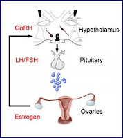
Various glomerulonephritides unfortunately often require treatment with chemotherapeutic agents, such as cyclophosphamide. A potential adverse effect of these agents in females of child-bearing age, is premature ovarian failure. Nate previously posted about this. Since then, there have been a couple of papers published that address this dilemma further.


A meta-analysis of six randomized controlled trials reported that GnRH treated women had a higher odds of return of spontaneous menstruation than those not treated (OR 3.46; 95% CI 1.13 – 10.57), but this did not translate into a significant difference in the rate of spontaneous pregnancy after chemotherapy. However, many people are cautious in the interpretation of these results, given the small sample size in the trials, heterogeneity of protocols and varying follow-up times.


More recently, a randomized trial of triptorelin vs standard care in women with breast cancer was published. This study, performed in Italy, randomized 133 women to GnRH and 148 to standard care. Subjects were reviewed one year after the last cycle of chemotherapy for occurrence of early menopause (defined as no resumption of menstrual bleeding and postmenopausal levels of FSH and estradiol one year after cessation of chemotherapy).
The rate of early menopause was 25.9% in standard care vs 8.9% in the GnRH group (p for difference <0.001).


Whilst encouraging, these findings must be tempered by the fact that recovery of menses does not directly translate to fertility (limited data was available for this cohort at the time of publication). Furthermore, the chemotherapeutic regimens used, dosing and the duration of treatment make it hard to generalize the findings to a ‘nephrology disease’ population. There are alternative methods available, with somewhat stronger recommendations from experts in the field – these include embryonic cryopreservation.

Friday, July 15, 2011

Calcium blocker induced dependent oedema


Dihydropyridine (DHP) calcium channel blockers are notoriously associated with dependent oedema, but what mechanisms underlie the pathogenesis of this finding? Here are some explanations from a nice review I came across.


Going back to Starling’s forces governing the formation of interstitial fluid – net capillary fluid filtration is influenced by hydrostatic pressure, oncotic pressure, capillary permeability and lymphatic drainage.

Upon change in posture from lying to standing, hydrostatic pressure in the vessels of the lower extremities increases. However, this is held in check by autoregulatory mechanisms which cause protective vasoconstriction via local sympathetic reflexes and myogenic reflexes – the latter are caused by smooth muscle contractions in response to increased vessel transmural pressure.

These myogenic reflexes are mediated in part by L-type DHP sensitive calcium channels. They allow calcium influx, causing pre-capillary smooth muscle constriction, thereby reducing hydrostatic pressure in the capillary bed – the final result is guarding against increased interstitial fluid accumulation.

DHP calcium channel blockers can prevent the entry of calcium to smooth muscle cells, preventing this protective pre-capillary vasoconstriction, facilitating the formation of dependent oedema. In patients with already impaired autoregulation (e.g. diabetics) calcium blockers may be associated with more profound leg oedema than others. Of course, there are likely other factors at play, including increased capillary permeability and other local neurohormonal influences.

Monday, July 11, 2011

Hypokalaemia induced polyuria

Last time we reviewed the proposed mechanisms by which hypercalcaemia can induce polyuria. It’s important to know that hypokalaemia can also precipitate this presentation. So here are the proposed mechanisms for this scenario.

Potassium is required for the thick ascending limb NaKCl2 co-transporter to operate. Remember that potassium is recycled via the apical ROMK channel, which facilitates ongoing sodium reabsorption in the TAL and maintenance of the medullary concentration gradient. Therefore, potassium deficiency may lead to concentrating defects via this pathway, leading to polyuria.
There is also some evidence that hypokalaemia leads to impaired responsiveness to ADH. The exact mechanism is unclear, but obviously decreased responsiveness to ADH may lead to large volumes of dilute urine.
Other interesting nuggets with hypokalaemia – movement of potassium out of cells in the proximal tubule (in the setting of hypokalaemia) is balanced by inward movement of hydrogen ions. The creation of an intracellular acidosis increases ammonia production by the proximal tubular cells, which may exacerbate problems in patients with decompensated liver disease.
Chronic potassium depletion (over a month at least) in humans can cause the development of vacuolar lesions in renal epithelial cells, primarily in the proximal tubule. If the deficiency persists over a longer time period, changes including interstitial nephritis, tubular atrophy and medullary cyst formation have been described. Potential mechanisms underlying these changes may be related to altered growth factor and cytokine production, or ammonia accumulation. This collection of findings has loosely been referred to as hypokalaemic nephropathy.

Sunday, June 26, 2011

Hypercalcaemia induced polyuria

So Leo’s post got me thinking about the pathophysiology of hypercalcaemia-induced polyuria. I wanted to share what I have learned in relation to the mechanisms of this phenomenon.


Calcium-sensing receptors (CaSR) are found on the basolateral membrane of the cells of the thick ascending limb of the loop of Henle. Binding of calcium, with subsequent receptor activation, appears to induce a downstream message to close off the luminal K channel. This in turn inhibits potassium recycling and thereby shuts off the NKCC2 cotransporter, leading to decreased reabsorption of sodium, potassium and chloride in this segment.


Following on from this, the medullary concentration gradient is diminished, leading to an inablility to concentrate urine and subsequent polyuria.


CaSR are also found on the luminal surface of cells in the inner medullary collecting duct. Activation of these, in the setting of increased distal calcium delivery, down-regulates AQP2 expression, again resulting in an inability to concentrate the urine.


Finally, there may be hypercalcaemia-induced PGE2 production in the thick ascending limb, which can further inhibit sodium chloride reabsorption.


Thankfully, the polyuria and concentrating defects associated with hypercalcaemia tend to regress with correction of the calcium levels. One caveat to this may be patients who have developed an interstitial nephritis (secondary to calcium deposition in the medulla ) from prolonged hypercalcaemia.

Sunday, June 12, 2011

Creatine vs Creatinine

Creatine monohydrate is a widely available dietary supplement. It frequently pops up during both outpatient and inpatient discussions on the differential for acute kidney injury. However, the literature is scant and appears to be conflicting. So, here are a selection of some of the papers that have been published on this subject.

Firstly, creatine itself is produced naturally in the body, mainly by the liver and kidney, and is transported mainly to muscle. It is involved in a complex interplay with phosphate to form phosphocreatine, facilitating a stable and rapid supply of energy on demand. Creatinine (our common, but imperfect, clinical surrogate of GFR) is a breakdown product of creatine.

One of the early reports of association of exogenous creatine with kidney damage was in 1998 - the case described acute on chronic renal impairment in a 25yo man on cyclosporine for FSGS, which was temporally related to ingestion of supplemental creatine (there was no reported change in proteinuria). Upon cessation of the supplement his serum creatinine returned to baseline. No biopsy was performed.


A second case reported biopsy proven acute interstitial nephritis in a 20yo man, which occurred four days after ingestion of creatine supplements. Again, upon cessation of creatine the renal function returned to baseline.


This review of the existing literature on creatine supplements, highlights the varying reported results of the effects of exogenous creatine on both CPK and serum creatinine levels, with some claiming no change and others claiming slight elevations. An interesting randomized, double-blind, placebo-controlled trial of 18 athletes looked at the effect of creatine ingestion (10 g/day over 3 months vs placebo) on cystatin C levels as an alternative measure of eGFR. The authors claimed that creatine caused no significant alteration in estimated GFR as assessed by cystatin C (in fact the cystatin C levels decreased).

Overall, it would appear that we are still awaiting more detailed information of potential renal risk associated with exogenous creatine use. The trials to date are relatively small and longer term follow up is needed. As ever, a detailed exposure history and close vigilance is required, especially in light of the current popularity of alternative and supplement based pharmacotherapy.

Tuesday, May 31, 2011

IgG-4 associated kidney disease

We heard a very interesting presentation today on autoimmune pancreatitis and concurrent systemic disease. The pathogenesis is felt to be related to IgG-4 producing plasma cells, and has now been linked to numerous systemic disease manifestations. The case in question had possible associated autoimmune hypophysitis, leading to presentation with a central diabetes insipidus picture.

These conditions first came to light following the description of autoimmune pancreatitis and recognition of the association of this disease with various other organ system involvement. The lesions in the affected areas had in common a dense infiltration of IgG-4 plasma cells and T lymphocytes. Involvement has been described in the liver, pancreas, gallbladder, kidneys, retroperitoneum and salivary glands as well as many other tissues.

In terms of renal involvement, the most commonly reported lesion is that of tubulointerstitial nephritis, again with a characteristic infiltration of IgG-4 positive plasma cells. Nodular lesions have also been described, which in many cases can be confusing for primary or secondary malignancies. Of note, sometimes these lesions can be FDG avid, and light up on PET scans, further confusing the picture with malignancy. Membranous nephropathy with IgG-4 positive deposits has also been described. Finally, retroperitoneal fibrosis may also predispose to obstructive nephropathy.

Diagnosis involves biopsy of the affected tissue with demonstration of IgG-4 positive plasma cells. Serum IgG-4 levels should also be requested and, if elevated, have a sensitivity for disease presence of ~70-95%.

Most successful reported treatments have been with corticosteroids, usually starting high and tapering to a lower maintenance dose. There is limited experience in relation to the duration of treatment with steroids or use of steroid-sparing agents in this condition.

Tuesday, May 10, 2011

Is lower salt intake bad for you – evidence?

An observational study of urinary sodium excretion (a surrogate of dietary salt intake) and it’s association with cardiovascular mortality was published in JAMA last week. The trial involved 3681 patients in Europe without cardiovascular disease at baseline. The details are below:


Type:
Observational cohort study comprising the primary Outcome cohort, from which two further follow-up cohorts were derived (BP cohort and HTN cohort):

- Outcome cohort (n=3681) – full set of baseline measurements available; used for mortality study
- BP cohort (n=1499) – full set of baseline and follow-up measurements; used for analysis of 24hr urine Na with BP
- HTN cohort (n=2096) – those without baseline HTN; used for analysis of incidence of HTN

Inclusion:
Participants of the FLEMENGHO and EPOGH studies

Exclusion:
Incomplete 24hr urine collections
Cardiovascular disease at baseline
At follow-up study date, further exclusions were based on losses to follow-up, incomplete 24hr urine collections, those taking anti-HTN medications, interval development of CV disease

Outcome:
Primary - Adjudicated all-cause and non-fatal and fatal cardiovascular outcomes
Others – incidence of HTN, X-sectional and longitudinal BP analysis
Results:
- Median follow-up 7.9 years; 47.3% male; 4.1% Diabetic; 28.4% smoke
- Mean 24hr urine sodium was 178mmol
- Adjusted all-cause mortality did not associate with urine Na excretion
- Adjusted CV mortality increased at the lowest tertile of urine Na excretion - adjusted HR for the lowest tertile of urine Na excretion 1.56 (95% CI 1.02, 2.36)
- Adjusted nonfatal CV events did not associate with urine Na excretion
- In adjusted analyses, a 100mmol increase in urine Na excretion associated with a 1.71mmHg increase in SBP, but no change in DBP
There have been a number of articles recently raising some eyebrows in relation to dietary salt intake and outcomes, particularly in diabetics. The article above is no exception, suggesting that those in the higher tertile of urine sodium excretion actually have less fatal CV outcomes during follow-up. The response from experts in the field has been swift and many concerns with the study have been raised.
Firstly, it’s observational – a major issue;
Second, there were only 84 CV-related deaths in the cohort of 3681 participants, making it hard to draw any firm conclusions due to lack of statistical power;
Third, there are concerns over the reliability of 24hr urine collections at the best of times, although they did measure 24hr urine creatinine excretion in an attempt to include only complete collections – unfortunately this automatically creates a certain type of selection bias, as only subjects with complete collections were evaluated – we don’t know anything about those who did not have complete collections;
Fourth, the baseline urinary Na was based on one single collections – an average of two or three would have been more robust.
It seems that extreme caution is required in interpreting these results – as ever, future research and randomized controlled trials have been called for - whether they happen or not is another matter.

Saturday, April 30, 2011

NKF Spring Clinical Meeting - Calcium reabsorption


Here are some points from one of the cases presented at the electrolyte workshop this afternoon:

The major teaching point was that the reabsorption of calcium closely follows that of sodium. Therefore, in sodium avid states, calcium tends to be reabsorbed also – conversely, in natriuretic states, calcium tends to be lost in the urine.

What about thiazides – they are associated with hypercalcaemia and hypocalciuria, but what is the mechanism there?
In the distal convoluted tubule, thiazides act by blocking the luminal Na/Cl cotransporter. There is also a luminal calcium channel called TRPV5, which allows calcium to be reabsorbed in the DCT via a transcellular route. Overall, this is felt to contribute around 15% of total tubular calcium reabsorption. It was thought that this channel might have played a role in the pathogenesis of thaizide-induced hypercalcaemia.


However, when they created a knock-out mice without TRPV5 and treated them with thiazides, hypercalcaemia still developed. So, it would appear that the mechanism simply involves decreased extracellular volume caused by the diuretics, leading to increased sodium reabsorption more proximally and consequent enhanced proximal calcium absorption. See this review.

Of note, the paracellular route for calcium reabsorption in the thick ascending limb is guarded by tight junctions between the cells. These are made up of proteins called Claudins. Genetic defects in Claudin proteins have been associated with impaired calcium reabsorption and subsequent hypercalciuria and stone formation, giving us some insight into the molecular level of calcium handling by the kidney.

This was a great meeting with lots of interesting discussions and talks. Roll on next year in Washington!

Thursday, April 28, 2011

NKF Spring Clinical Meeting - BP in CKD

I attended a very interesting talk from Dr Bakris today on the latest approaches to BP targets in the CKD population. The highlights are nicely illustrated in a systematic review published in the Annals this year.

They searched for randomized controlled trials comparing blood pressure targets in adults with CKD that had more than 50 participants per group; at least 1-year follow-up; with death, kidney failure, cardiovascular events, change in kidney function, number of antihypertensive agents and adverse events as the outcomes of interest. There were three trials that met these criteria, with a total of 2272 subjects contributing data.

Interestingly they found that there was no evidence to suggest that a target of less than 125/75 to 130/80 mm Hg was more beneficial than targets less than 140/90 mmHg.

However, the degree of proteinuria may be an effect modifier here – in this regard they found lower quality evidence to suggest that in those with macroalbuminuria (0.3 – 1.0g/day), the lower target may be beneficial.

The main limitation of this review was that the trials involved were limited to non-diabetic CKD patients.

The tide of opinion may be turning in relation to BP guidelines, so watch closely for any announcements from the major societies in relation to this. It’s important to avoid complacency though, and a relaxation of the target shouldn’t mean we place less emphasis on BP control.

Wednesday, April 27, 2011

NKF Spring Clinical Meeting - IgA

The NKF Spring Clinical meeting started today in Las Vegas. I thought I’d share of the interesting highlights from some of the talks I’ve attended. This post relates to Dr Appel’s lecture on IgA nephropathy, where he described the recent literature on pathogenesis and treatment and a relatively new classification system called the Oxford-MEST system.


This classification system was devised at an expert group meeting in England. Pathologists (blinded) retrospectively examined 265 biopsies from adult and paediatric patients with IgA, who had five-year follow up data available. The biopsies were scored on pathological variables and entered into a prediction system based on the follow-up data available. They came up with four features that were strongly predictive of renal outcomes:


M – mesangial hypercellularity; scored 0 or 1
E – endocapillary proliferation; scored 0 or 1
S – segmental glomerulosclerosis; scored 0 or 1
T – tubular atrophy/interstitial fibrosis; scored 0, 1 or 2


A notable absence in this classification system is presence of crescents – the reason why this is not included is simply that in the 265 samples they analyzed, there were too few samples with crescents to make any significant inference about.


Further validation of this score is ongoing in various samples, but it appears to be gaining recognition and popularity and is something we should be aware of.

Monday, April 25, 2011

Thirst and xerostomia

One of the major problems we struggle with in haemodialysis patients is excess, volume, inter-dialytic weight gain and ultrafiltration. Despite constant reminders by nursing staff, physicians and nutritionists, patients seem to find it extremely hard to restrict their fluid intake to what we’d like it to be.


The reasons for this are variable and include social and cultural differences, habit, psychological diagnoses, medications and likely the very dialysis procedure itself by means of salt-loading. One of the clinical manifestations that is important to differentiate from thirst (a central mechanism driven by tonicity) is xerostomia (dry-mouth).


Xerostomia has a broad differential of possible causes and includes medications (particularly those with anti-cholinergic effects), anxiety, radiation therapy, connective tissue disease and rarely, autonomic disease. It is important to examine the long list of medications prescribed to our patients for possible culprits contributing to dry mouth and the excessive fluid intake that results from it.


I came across a small study of 43 HD patients followed for two years in Holland (not the most robust study, but interesting nonetheless). Unstimulated and chewing-stimulated 5 minute saliva collections were performed at baseline and at 2 years, along with a formal xerostomia inventory and thirst questionnaires. Then a formal dental examination was performed on each patient examining for decay, oral hygiene and periodontal disease.


Twenty patients were transplanted during follow-up. The most interesting finding was that those who were transplanted experienced an increase in salivary flow rate from 0.3ml/min to 0.44ml/min (p=0.002) and a decrease in thirst score from 10.6 to 8.1 (p=0.02); there were no significant changes over the 2 years for those who remained on dialysis. Also, there were no detectable differences in oral health between the 2 groups and the 2 time-points.


It’s possible that there is something peculiar about the state of ESKD that alters salivation and contributes to the symptom of dry mouth (supported by the observation of improvement after renal transplantation). Perhaps attention to the medication history and trying to distinguish the symptoms of thirst from dry-mouth may help us to help our patients in their ongoing battle with inter-dialytic weight gain.