Monday, August 31, 2009

General Rules Regarding the Dialyzability of Drugs

For many common drugs, we have a good idea of whether or not they are dialyzed off. We know that it's important to re-dose vancomycin after a dialysis session since some of it is removed; conversely, drugs such as ketoconazole are essentially non-dialyzable. However, situations may arise--for example, in instances of a drug overdose--where the nephrologist may get a call asking to perform emergent dialysis in order to remove a much less common medication. How does one decide whether a dialysis procedure would be effective or not?
There are a few basic rules that govern whether a drug is dialyzable or not. Not surprisingly, size matters. Molecules with a low molecular weight (e.g., less than 500 Daltons) are easily cleared, whereas larger molecular weight compounds (e.g., greater than 2000 Daltons) are not. However, just because a molecule has a low molecular weight does not necessarily mean that it is efficiently cleared. A good example is Lithium, (atomic #3 on the periodic table) which has a fairly high volume of distribution, and therefore longer (and sometimes multiple) dialysis sessions are necessary in order to effectively dialyze an invidual with Lithium intoxication. A general rule is that drugs with a VOD greater than 1Liter/kg are poorly dialyzed. In a related vein, drugs with high protein binding or which are highly lipophilic are also poorly dialyzed.

As an example, let's take vancomycin. It has a molecular weight of 1486 Daltons with a VOD of between 0.5-0.9 Liters/kg. Therefore, one would anticipate based on these rules that it is at least partially removed by dialysis. These principles can be used to roughly assess the dialyzability of various unknown compounds as well.
Bonus points for being able to visualize the stereogram of vancomycin below!

Saturday, August 29, 2009

Renal TB

TB is classically thought of as a pulmonary disease--and in general, this is true, as pulmonary TB is by far the most common (and often very severe) manifestation of infection by Mycobacterium tuberculosis. However, the most common NON-pulmonary manifestation of TB worldwide is infection of the genitourinary tract, accounting for about 27% of non-pulmonary TB in most series according to this 2001 JASN review.

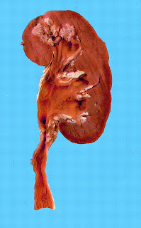
The name "renal tuberculosis" is a little misleading, since TB infection can actually cause renal problems by a variety of different mechanisms. Renal involvement may occur as part of a constellation of symptoms with miliary TB, in which there is mycobacterial "bacteremia/septicemia". The preferred site for mycobacteria in the kidney is the renal medulla, where caseating granuloma can lead to necrosis and tissue destruction, occasionally resulting in papillary necrosis. Large caseating necrotic areas can lead to a clinical picture very similar to pyelonephritis. Alternatively, infection in the lower urinary tract can lead to anatomic abnormalities, such as ureteric strictures, which over time can progress to frank obstruction and even ESRD. In addition to these more "mechanical" explanations for renal failure, there have been several reports that chronic tuberculosis infection is a potential cause for tubulointerstitial nephritis. In India, where TB is very common, AA amyloidosis secondary to the chronic inflammatory state induced by TB infection is a well-recognized cause of kidney damage.

Genitourinary TB may be diagnosed by culture or PCR of the organism from the urine (or sputum, if there is pulmonary involvement). Detecting acid-fast bacilli from the urine is another possibility though apparently this is less specific than culture or PCR as there are some environmental mycobacterial species which may colonize the lower urinary tract. Renal TB may be suspected if the patient has signs and symptoms of a urinary tract infection with a workup that demonstrates pyuria but a negative urine culture. Obviously, travel/occupational history plays an important role in identifying individuals who are at higher risk for TB. Another clue to the diagnosis of renal TB is hypercalcemia: instead of being mildly hypocalcemic like most advanced CKD patients, patients with TB infection may have high levels of 1,25(OH) vitamin D as a result of granulomas having the ability to synthesize active vitamin D, similar to the situation in sarcoidosis. In the U.S., a very low percentage of patients (.004%) list TB as a cause for their ESRD, but in some European countries (e.g. Greece) the percentage is as high as 5%, and likely much much higher than this in many African countries. It is important to recognize in that TB is very much a treatable disease (and therefore a preventable cause of CKD/ESRD).

Thursday, August 27, 2009

Basic Review: The Renin-Angiotensin-Aldosterone Axis


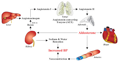
One of the coolest aspects of the renin-angiotensin-aldosterone system (RAAS) is that it involves multiple organ systems: the liver, lung, adrenal gland, kidney, and vasculature are all prominently involved. It never hurts to review basic physiologic principles, right? Listed are the three main components of the RAAS and their main mechanisms of action.

1. Renin is a peptide hormone secreted from the juxtaglomerular cells of the afferent arteriole in response to 3 main stimuli: (a) renal hypoperfusion, (b) decreased distal chloride delivery to the macula densa, and (c) increased sympathetic activity. Renin antagonists such as aliskiren are presently being tested as antihypertensive agenst with thus far promising results.

2. Angiotensinogen--which is synthesized and secreted from the liver--is cleaved by renin in the systemic circulation to form angiotensin I.

Angiotensin I is cleaved to form angiotensin II by angiotensin converting enzyme (ACE), which is found predominantly within lung endothelium. ACE-inhibitors, as their name implies, targets the ACE enzyme and is one of the most potent anti-hypertensives (and GFR-preserving) therapies available.

Angiotensin II has the following physiologic effects, which it carries out via binding to AT1 and AT2 receptors. Drugs which block the ability of angiotensin II to bind to its receptors ("angiotensin receptors blockers", or ARBs) make up another highly successful and renoprotective antihypertensive therapy. Angiotensin II binding to its receptors have the following major effects:

a) angiotensin II acts as a systemic vasoconstrictor.
b) angiotensin II causes renal efferent arterial vasoconstriction. Acutely, efferent vasoconstriction should increase GFR; however, over time the increased glomerular pressure leads to glomerular damage and, ultimately, renal injury.
c) angiotensin II increases secretion of aldosterone from the zona glomerulosa of adrenal cortex.

3. Aldosterone: in cortical collecting duct cells, aldosterone diffuses into the cell and interacts with the mineralocorticoid receptor, which upon binding translocates to the nucleus and increases expression of ENac. The end result of aldosterone action is sodium reabsorption and potassium & hydrogen secretion. In addition to angiotensin II, hyperkalemia can also stimulate aldosterone secretion. The drug spironolactone interferes with aldosterone interacting with its receptors, and can be effective in the treatment of hypertension.

Wednesday, August 26, 2009

Bundling

It's a hot topic amongst dialysis centers around the country: bundling. Here's my attempt to explain it in a brief manner. If anybody has corrections or comments about this explanation, feel free to make them known.

With the Social Security Act of 1973, all U.S. ESRD patients were given the right to free dialysis--and since that time, the ESRD Program has comprised a very significant portion of the overall Medicare Budget. The Centers for Medicare and Medicaid Services (CMS), a division of the Department of Health and Human Services (HHS), is in charge of administering the ESRD Program and setting the reimbursement prices for dialysis centers.

Currently, CMS divides the ESRD budget into two distinct services. First, there is a "bundled" payment which takes into account all the nursing, dialysis equipment, and many of the routine lab tests; essentially, one lump sum per patient per dialysis treatment is given which does not vary. Second, there is a category of "separately billable" items which includes the injected medications administered during dialysis and a few of the more recent lab tests. According to the current plan, each "separately billable" item is reimbursed at a rate 6% above the manufacturers' average sales price (ASP). Dialysis centers can make a profit from this policy, and accordingly, the more "separately billable" items are ordered, the greater the ability to generate profit. This has been especially welcomed by dialysis units, as the bundled payment has not been adjusted for inflation and the units rely more and more heavily on "separately billable" items to cover the increasing costs of running a center.

There is now a strong push by CMS to change the ESRD policy such that the entire dialysis process--both the dialysis treatment itself as well as any injectable medications or labs--become a single "bundled payment." According to this report by the Government Accountability Office (GAO), a single bundled dialysis care payment would "increase efficiency and clinical flexibility."

The medication Epogen is central to understanding the current move towards a bundled payment. Epogen was by far the most pricey "separately billable item" within the ESRD budget, accounting for about $2 billion annually. In the report, the fact that Amgen (the company which makes Epogen) has no competition on the market is cited as a reason for these staggering costs. The report goes on to suggest that placing Epogen within a bundled payment would force physicians to use the drug more judiciously and allow them to adapt strategies that could minimize Epogen doses--for example, subcutaneous rather than intravenous dosing.

The proposal of bundled dialysis payments is, understandably, still a controversial one. Will dialysis units be fairly compensated in a bundled system? We don't have the proposed numbers yet to judge. Will patients still have access to important drugs and breakthroughs? Some have even suggested that a bundling policy might discourage some dialysis centers from taking African-American patients, who tend to have higher Epogen requirements, and therefore lead to racial discrimination; medically complex patients might also fall into the category of an "undesirable dialysis patient" from a cost perspective. The one thing that is for sure is that whatever the policy turns out to be, it will undoubtedly have major effects on nearly every nephrologist practicing with the U.S.

Tuesday, August 25, 2009

Pathology of BK Nephropathy

BK Nephropathy refers to damage suffered to a renal allograft as a result of the polyomavirus BK virus.   It is named based on the fact that it was originally isolated in the urine of a kidney transplant patient with the initials "B.K." in 1971.  Infection with BK virus rarely causes problems in immunocompetent individuals; however, the increased use of potent immunosuppressives such as tacrolimus and MMF have increased the risk of BK nephropathy.

While BK nephropathy is often suspected based on monitoring of either serum or urine BK virus levels, the allograft biopsy is presently an essential aspect of its diagnosis.  A few of the key characteristics of BK nephropathy worth noting:

1.  interstitial fibrosis:  there is a staging system for BK Nephropathy (see this article)--it lists three stages (A, B, and C) which vary according to the degree of interstitial fibrosis and, not surprisingly, is associated with prognosis. 

2.  intranuclear inclusion bodies:  renal epithelial cells may contain large nuclei indicative of viral infection.  Viral infection may also occur in transitional epithelium of the bladder, and when shed into the urine show up as decoy cells, another non-invasive means of helping obtain a diagnosis of BK Nephropathy.

3.  Immunohistochemical staining against the SV-40 T-antigen.  These special stains will often clinch the diagnosis of BK Nephropathy, as it is highly specific for all polymaviruses (the most common of which is BK virus, but on occasion is JC virus).  Staining should be restricted to the nucleus and is a sign of active viral replication.

4.  Electron microscopy demonstrating viral particles:  the viral particles are typically intranuclear, 30-50nm in diameter, and have a icosahedral capsid structure (see below).

Monday, August 24, 2009

Recent Urine NGAL Biomarkers Studies

Two recent articles in JASN--one by Siew et al and the other by Paragas et al--provide further support to the idea of using urinary NGAL as a biomarker for acute kidney injury. An accompanying editorial by Lynda Szczech, entitled, "The Development of Urinary Biomarkers for Kidney Disease Is the Search for Our Renal Troponin", provides a thoughtful analogy for thinking about how best to use these tests. The author emphasizes that biomarkers will not necessarily replace creatinine and urine output as a means of assessing AKI--but rather it will supplement these more traditional tests, much like troponin is now used in conjunction with older methods (e.g., EKG analysis) is diagnosing myocardial injury. A marker such as urinary NGAL may be a better marker for injury, as serum creatinine is really a marker of kidney function, and becomes elevated far after the kidney insult.

In the study by Siew et al, over 400 critically ill patients underwent urinary NGAL measurement within 24 hours of admission to an ICU; these patients were then followed prospectively and assessed for AKI, as defined by an increase in serum creatinine of greater than 0.3mg/dL or a greater than 50% increase in the baseline creatinine. The investigators found that elevated urinary NGAL levels was moderately successful in predicting AKI.

In the second study by Paragas et al, investigators looked at the ability of urinary NGAL to distinguish between HIV patients with a collapsing FSGS pathology (e.g., "HIVAN") compared to HIV patients that had either normal kidney function or CKD from another cause. Importantly, patients with HIVAN had 11-fold higher urinary NGAL levels compared to HIV-positive controls without a reduced GFR, and still 5.5-fold higher urinary NGAL levels compared to HIV-positive controls with CKD due to a cause other than HIVAN. These findings may prove useful in terms of diagnosing patients with HIV and rapidly declining renal function with HIVAN in a non-invasive manner (e.g., no biopsy). While biopsy should still likely remain the gold standard until these findings can be confirmed with a larger n, it could potentially be useful information in patients where biopsy is deemed too risky to proceed--a situation in which HIVAN patients may commonly find themselves.

Friday, August 21, 2009

pre-dilutional versus post-dilutional CVVH

There are two general strategies for CVVH replacement solution entering into the blood circuit: pre-dilution (in which replacement solution is mixed with the blood prior to its entry into the filter), or post-dilution (in which replacement solution is mixed with the blood after it has passed through the filter). Each method has its own advantages and disadvantages. Where I have trained, we always use the pre-dilution method. The theoretical advantage here is that because the blood is diluted prior to entry into the filter, there is a lower risk of clotting the filter, allowing for longer filter life and reduced down-time for the CVVH machine. We often claim that CVVH is a continuous therapy, but due to technical issues it is not uncommon for the therapy to be halted for several hours out of a day; pre-dilution technique may help minimize this. Furthermore, pre-dilution allows for near-limitless ultrafiltration rates, essential when rapid volume removal is necessary. The main cited drawback of the pre-dilution method is that much of the fluid passing through the filter is actually replacement fluid--thereby leading to a reduced efficiency of about 10-15% in clearance when compared to tost post-dilutional method, in which all the fluid moving through the filter is the patient's. Generally this is not a problem given that this is a near-continuous therapy, but proponents of the post-dilution method claim improved efficiency as an advantage. It is important to specify pre- versus post- dilution methods in the interpretation of various publications in CVVH.

Thursday, August 20, 2009

Rule of Thirds in Membranous Nephropathy

Keeping with our recent theme of "numerical rules in Nephrology", let's briefly discuss the "Rule of Thirds" for Membranous Nephropathy.

The "Rule of Thirds" is frequently taught in medical school and a useful way of thinking about the prognosis of membranous nephropathy. It states that roughly speaking, all patients with the diagnosis of membranous nephropathy can be broken down into 3 equally populated groups with different prognoses:

One third of the patients will experience a spontaneous remission (this particular group, while certainly a desired outcome for all patients, has made interpretation of clinical trials in membranous nephropathy somewhat difficult--you don't know if a particular test subject has gotten better because of a treatment, or would have done so on their own).

One third of the patients will have persistent proteinuria and a reduced but stable renal function.

One third of the patients will have progressive loss of GFR, often leading to ESRD. According to this source, the overall incidence of ESRD is 14% at 5 years, 35% at 10 years, and 41% at 15 years.

While useful for a global understanding of membranous nephropathy, however, the Rule of Thirds is a little too simplistic--we can more accurately predict a patient's prognosis based on risk-stratification. In this 2005 JASN review by Cattan, the author presents a well-utilized strategy for the decision of how aggressively to treat patients with membranous nephropathy. Generally, it advises breaking patients up into low-risk, medium-risk, and high-risk groups based on their response to conservative therapy (e.g., ACE-I/ARBs, edema and lipid management, blood pressure control, etc) over a 6-month period.

Those in the low-risk group (normal renal function and less than 4 grams/day proteinuria over a 6-month period) have less than a 5% risk for progression over a 5-year period and therefore should not be given aggressive immunotherapy.

Those in the medium-risk group (normal renal function and persistent proteinuria between 4 and 8 grams/day proteinuria over a 6-month period). This group has a much higher risk of poor outcomes without treatment than the standard "Rule of Thirds" would suggest, and therefore the data would suggest treatment such as cycling of cytotoxic drugs and steroids.

Those in the high-risk group (worsening renal function and proteinuria greater than 8 grams/day over a 6-month period) represented only about 10% of all patients with idiopathic membranous nephropathy, but make up a very high percentage of those who eventually go on to ESRD. These patients should receive aggressive therapy with the possibility of newer protocols (Rituxan, cyclosporine for failure to achieve remission, enrollment in new trials) and be advised that dialysis is a very real eventual possibility.

Wednesday, August 19, 2009

DDx for Elevated BUN or Cr without Low GFR

Most of the time when we are consulted for an inpatient with a rising creatinine, the assumption is that the kidney is to blame. However, there is an important list of non-renal conditions which can increase either BUN or creatinine independently of low GFR.

An elevated BUN without AKI can occur in the following situations:
1. G.I. bleeding (because digested blood is a source of urea nitrogen).
2. excessive protein intake or a catabolic state.
3. steroid use.
4. TPN.
5. multiple boluses of iv albumin given.

An elevated creatinine (structure pictured above) without AKI can also occur, though less commonly than an elevated BUN without AKI. Recall that a percentage of creatinine clearance occurs via secretion; therefore, drugs which inhibit creatinine secretion such as Bactrim, probenecid, or Pepcid can increase serum creatinine without actually affecting the GFR. Furthermore, intense exercise can transiently increase the creatinine. Finally, some chemicals (e.g., nitromethane, used as a component of types of fuels and solvents) can interfere with the assay by which creatinine is detected in the laboratory.

Tuesday, August 18, 2009

Bence-Jones Protein

I had always assumed that the "Bence-Jones" protein--essentially, the demonstration of monoclonal light chains on urine protein electropheresis (UPEP)--was named after two doctors, Bence & Jones.  However I recently found out that actually it was named after a single individual:  Henry Bence Jones, a famous British physician and chemist.  In 1848, he was cited as the driving force for the investigation of an unusual chemical analysis discovered in the urine of a patient with myeloma in a paper titled "On the microscopical character of mollities ossium" (mollities ossium was the name for myeloma, which at the time was thought of as a bone disease based on the osteolytic bone metastases which resulted).  In this paper, he described the appearance of a precipitate which occurred when the urine was heated to 50-60 degrees, disappeared when boiled, and reappeared again when the urine cooled--this substance is now known to be the same urine light chains which result in cast nephropathy.

According to this brief biography of Henry Bence Jones, he published on a variety of topics including renal calculi and gout, and was an early proponent of the urinalysis (both urine microscopy as well as chemical analysis of the urine) in diagnosis.  He was also apparently the physician for Charles Darwin, and published the then-definitive biography of the physicist Michael Faraday.   

Monday, August 17, 2009

the von Kossa Stain for Acute Phosphate Nephropathy

One of the hot topics in Nephrology over the past few years has been the epidemiologic and histopathologic studies suggesting oral sodium phosphate colonoscopy preparations as a cause of acute phosphate nephropathy. As evidence continued to mount, the FDA in December 2008 forbade the over-the-counter sale of oral sodium phosphate products; however, they are still available by prescription under the names "Visicol" and "Osmoprep." These preps are still favored by many gastroenterologists (and patients) based on the fact that it is much easier to take than the more traditional polyethylene glycol-based colonoscopy prep (e.g., "Go-Lytely").

Biopsies of patients with acute phosphate nephropathy tend to show abundant calcium phosphate crystal deposition, mostly within the distal convoluted tubules and collecting ducts, but sometimes also in the interstitium. A good detection method is the use of the von Kossa stain, which stains certain calcium-containing salts such as calcium phosphate a brownish-blackish color.

Sunday, August 16, 2009

Rule of 4s for Diagnosing Ultrafiltration Failure in PD

In peritoneal dialysis, ultrafiltration failure is defined as the inability of the peritoneal dialysate to maintain euvolemia--and generally speaking, this implies some degree of hypervolemia. Technically, it can occasionally result from trivial explanations--for example, an incorrect PD prescription not properly calibrated to that patient's peritoneal equilibration test, or poor patient compliance with an appropriate PD regimen. But usually when the term ultrafiltration failure is used, it often implies that the peritoneal membrane has evolved into such a high transporter status that net ultrafiltration becomes impossible.

One useful definition for ultrafiltration failure status, based on a simple office test, is the following
"Rule of 4's": give the patient a bag of 4.25% dextrose-based PD fluid for 4 hours. If there is not net ultrafiltration of greater than 400 mL over this time period, then the patient has ultrafiltration failure. Ordinarily, PD with 4.25% solution is a reliable way of removing excess fluid, to the point where in the pre-CVVH and inpatient HD days, installing a PD catheter and initating exchanges with 4.25% solution was actually a means of treating acute inpatient pulmonary edema in the ICU.

Ultrafiltration failure often correlates with the loss of residual renal function and the onset of anuria. Strategies to avoid UF failure include using icodextrin periodically for longer dwells, minimizing salt intake in the diet, and using a cycler overnight. If these maneuvers do not succeed, ultrafiltration failure may ultimately signify an inability to proceed with PD and the necessity of transitioning to hemodialysis.

Thursday, August 13, 2009

Rule of 6s for Dialysis Access Placement

The "Rule of 6s" describes an easy way to evaluate the maturity of a recently-placed arteriovenous fistula for dialysis access--and is even mentioned by name in the most recent KDOQI Guidelines for Dialysis Access.  The Rule of 6s is as follows:

6 weeks after the AV fistula has been placed, the fistula should:
(a) be able to support a blood flow of 600 ml/min.
(b) be at a maximum of 6mm from the surface.
(c) have a diameter greater than 6mm.

Failure to achieve these goals warrants a further investigation, usually in collaboration with the access surgeon who placed the fistula, into why the fistula did not mature.  Ideally, the existing fistula can still be encouraged or modified to eventually achieve maturity; if not, a new access site can be attempted.  

Wednesday, August 12, 2009

The Four Types of Immune Sensitivity Reactions & Renal Examples for Each

Basic Clinical Immunology:  there are four types of hypersensitivity reactions.  Below is a listing of all four types and, since this is a Nephrology Blog, renal examples for each one.

Type I Immune Reaction (anaphylactic):  Type I hypersensitivity reactions are IgE-mediated responses which occur relatively rapidly following exposure to a previously-encountered antigen.  A classic example of this in nephrology is the "Type A" dialyzer reaction which can manifest as acute hypotension within minutes of beginning dialysis.  Classically, this has been due to treatment of dialyzer cartridges with ethylene oxide, and fortunately this type of reaction is much less common today than previously.  

Type II Immune Reaction (antibody-mediated):  Type II immune reactions are caused by antibodies which react with self-antigens, and the direct toxicity which results in this interaction.  Two good renal examples are Goodpasture's Syndrome (in which an antibody against an epitope in the glomerular basement membrane results in rapidly progressive glomerulonephritis) and membranous nephropathy (see my recent post on the exciting discovery of an antibody against phospholipase A2 receptor, a podocyte antigen, thereby causing proteinuria).

Type III Immune Reaction (immune complex-mediated):  Type III immune reactions are also antibody-mediated, but they involve the deposition of pre-formed immune complexes and resultant complement activation.  Immune complexes GN's such as lupus nephritis and post-strep glomerulonephritis are the classic examples here. 

Type IV Reaction (delayed hypersensitivity):  Type IV immune reactions are mediated predominantly by T-cells, which explains why the lag time from exposure to immune response is somewhat delayed compared to the other immune reaction types.  A good illustrative example from the field of transplant nephrology is the acute cellular rejection response that occurs within a few days of a renal transplant.  Such reactions are best managed by agents which diminish T-cell function (e.g., calcineurin inhibitors, thymoglobulin, etc).  

Tuesday, August 11, 2009

Amazing Dialysis Biker

Wow. In case you hadn't heard the news, Shad Ireland, a 37 year-old dialysis patient, recently completed a cross-country bike trip which involved cycling from California to Washington, DC. Mr. Ireland has been on dialysis for 27 years, and had to plan his route so as to receive his regularly-scheduled dialysis treatments. He has also apparently completed an Iron Man Triathlon. His bike trip was meant to raise awareness for CKD/ESRD and certainly seems to have succeeded; the completion of his journey has been well-documented by several major news stations.

Monday, August 10, 2009

Lipid-Lowering Effect of Renagel

This has been known for awhile, but it is worth repeating--sevelamer (Renagel), in addition to its ability to act as a phosphate binder, also results in LDL cholesterol-lowering effects.

Two of the early articles (a 1998 Clinical Nephrology study by Wilkes et al, as well as this 1998 NDT paper by Goldberg et al), first noted this unexpected association.  In the former paper, a group of dialysis patients subjected to sevelamer hydrochloride therapy following a phos-binder washout period was noted to have a 35.9% fall in LDL cholesterol with an HDL cholesterol level that remained relatively stable.  This LDL-lowering effect is also present in the newer formulation of Renagel, sevelamer carbonate.  

It is unclear exactly what this lipid-lowering effect may indicate, especially considering the repeated finding that statins appear to be less efficacious in CKD/ESRD patients than in the general population in terms of lowering cardiovascular mortality.  Still, it is tempting to speculate that cholesterol-lowering should be a good thing for CKD/ESRD patients given their high rate of CVD.  The jury is still out in terms of what is the optimal phosphate binder to use in a given clinical scenario, but the lipid-lowering effect of Renagel does make it somewhat unique from the other phosphate binders available.  

Saturday, August 8, 2009

When To Refer For Dialysis Access?

Despite the knowledge that AV fistulas and grafts give superior outcomes when compared to dialysis catheters, there remain a large chunk of patients who begin dialysis without a fistula or graft. Conversely, there are also examples of "unnecessary procedures"--individuals with advanced CKD who get an AV fistula placed, but never use it either because their CKD never progresses to ESRD or because they die before reaching dialysis. These facts underline the difficulty nephrologists have with accurately predicting the progression of CKD to ESRD.

How should one decide precisely WHEN to refer for dialysis access? KDOQI does have guidelines--they state that individuals with CKD should have a fistula placed once the GFR is less than 25, the creatinine is greater than 4 mg/dL, or there is the anticipated need of dialysis within 1 year--but these are largely based on expert opinion.

One paper which seeks to address this issue in a more quantiative manner is a 2007 KI paper by O'Hare et al. Briefly, investigators looked at a large retrospective cohort of patients with an eGFR less than 25 calculated the ratio of unnecessary to necessary surgeries. An unnecessary surgery, for instance, was one in which a fistula or graft was placed but dialysis was not required within the 1-year period--either because their CKD remained stable or because they died before reaching ESRD.
In all the hypothetical scenarios they examined, older patients would have been more likely than younger patients to receive unnecessary procedures. For instance, if all patients were given a fistula at the time of cohort entry (eGFR less than 25), the ratio of unnecessary to necessary procedures would have been 5:1 for patients aged 85-100 years, but only 0.5:1 for those aged 18-44 years.

The bottom line: the decision as to timing of dialysis access placement should probably take into account age: young patients with a low GFR are more likely to eventually reach ESRD because of their longer predicted lifespan, and thus a case could be made to be more aggressive with access patient in this group. Obviously, every patient is unique and the nephrologist must take into account multiple issues (e.g., rapidity of CKD progression, other comorbidities, etc) in making this important decision.

Friday, August 7, 2009

iv iron preparations

Because of the generally poor GI absorption of iron in the setting of ESRD, iron supplementation in dialysis patients is now carried out by intravenous formulations of iron complexed to various carbohydrates. The idea is that these carbohydrate moieties can function as "molecular shields", allowing for the safe delivery of iron to its target tisues while simultaneously preventing iron-mediated oxidative damage. Here are some of the main iv iron formulations and their unique attributes:

1. iron gluconate (Ferrlicit). In my limited experience, it appears to me that Ferrlicit and Venofer control the lion's share of iv iron formulations in U.S. dialysis centers. A typical course of Ferrlicit typically given in the ESRD patient is 125mg iv qdialysis session x 8 doses.

2. iron sucrose (Venofer). Also a popular option, the typical dosing for Venofer is 100mg iv qdialysis x 10 doses. Both Venofer and Ferrlicit offer fairly rapid release of iron. Also, Venofer is FDA-approved for iron repletion in non-dialysis-dependent CKD patients whereas Ferrlicit is not.

3. iron dextran (Dexferrum, Imferon): this is not used much anymore because of a significantly higher risk of anaphylactic reactions than the more modern Ferrlicit and Venofer. Iron dextran was typically given as a smaller "test dose" prior to giving the full dose, as a precaution against anaphylaxis.

4. low-molecular weight iron dextran (CosmoFer, InFed): it is important to distinguish low-molecular weight dextran from high molecular weight dextran because its risk of adverse events is so much lower.

5. ferumoxytol (Feraheme): this is a newly-released formulation of "iron oxide nanoparticles." Sounds very space-age, doesn't it? The reported advantage is that it can be given in large bolus doses--thus making it preferable for the treatment of iron deficiency in CKD, where a patient would otherwise be required to make multiple trips to an infusion center to get their Venofer or Ferrlicit. Further assessments of safety and efficacy are still needed.

Thursday, August 6, 2009

Lifeport Kidney Transporter

Here's a cool little feature from the folks at CBS News describing the use of the "Lifeport Kidney Transporter"--a new hypothermia-perfusion device which functions as a significant upgrade from the standard cooler in which donor kidneys are transported from place to place.

A 2009 NEJM article by Moers et al actually showed in a randomized controlled trial that the Lifeport Kidney Transporter resulted in a superior one-year allograft survival compared to the cooler method. The control & experimental groups were pretty evenly matched in that each of the two kidneys from a given cadaveric harvest was assigned to opposite groups, which is a nice experimental design.

Wednesday, August 5, 2009

Development of CKD in Non-Renal Organ Transplants

As the longevity of non-renal organ transplants increases, so too does the risk of eventually getting chronic kidney disease. The occurrence of post-transplant CKD is relatively common due to two primary factors: (1) a dependence on calcineurin inhibitors for immunosuppression, which over the longhaul can result in chronic nephrotoxicity, and (2) an increased susceptibility to repeated bouts of acute kidney injury in the peri- and post-transplant state, often due to hemodynamic fluctuations such as hepatorenal syndrome (in those with liver failure) or cardiorenal syndrome (in those with heart failure).

Not surprisingly, different organ transplants are associated with different rates of CKD and ESRD than others. In a paper by Ojo et al in NEJM in 2003, investigators determined which organ transplants had the highest rate of renal failure. Topping the list was intestine transplants, which occurred in a whopping 21.3% of recipients over a 5-year period. This was followed by (in decreasing order) liver transplants, lung transplants, heart transplants and a much more modest risk of about 6.9% for those with a combined heart-lung transplant.

Not surprisingly, key predictors of CKD in this patient population in a multivariate analysis included old age, female sex, pretransplantation hepatitis C infection, hypertension, diabetes, and the presence of post-op AKI.

Although not mentioned in this particular paper, the prevalence of CKD/ESRD associated with stem cell transplants is overall lower than for the solid organs listed above. One potential reason for this is that calcineurin inhibitors are not required chronically in many of these patients.

Tuesday, August 4, 2009

New Urine Test for Appendicitis

Traditionally surgeons have relied on a well-honed abdominal exam, combined with abdominal imaging usually in the form of an iv-contrast CT scan, to make the diagnosis of acute appendicitis. Nevertheless, there remain significant instances of "false negatives" (usually a delay in diagnosis due to real episodes of appendicitis not picked up on imaging, occasionally resulting in appendiceal rupture) as well as instances of "false positives" (individuals who undergo appendectomy due to suspected appendicitis, but turn out to have no local inflammation on biopsy).

In order to improve the rapidity of diagnosis of appendicitis, Kentsis et al sought to identify new urine biomarkers which could conceivably help, as described in a recent issue of Annals of Emergency Medicine. In brief, their study subjected 12 urine specimens--6 from patients with appendicitis and 6 from patients without appendicitis--to mass spectrometry. They identified a list of several potential biomarkers which were elevated uniquely in appendicitis. They then attempted to validate selected markers in 67 children admitted for suspected appendicitis, 25 of which eventually turned out to have true appendicitis. The most promising candidate was leucine-rich alpha-2-glycoprotein (LRG), which demonstrated near-perfect sensitivity & specificity for predicting appendicitis early on.

I think the study is relevant to nephrology to the extent that substances specific to disease processes other than the kidney can be detected in the urine: "the urine is the window to the soul." It also is another example of how this cool "high-throughput"-type technology will continue to lead to the identification of new useful diagnostic tests relatively quickly.

Monday, August 3, 2009

Serum Alkaline Phosphatase as risk factor for CV Disease in ESRD Patients

Quick one today: interesting article in a recent issue of C-JASN by Shantouf et al that demonstrates a link between serum alkaline phosphatase levels and vascular calcification in patients with ESRD. Briefly, the study examined 137 randomly-selected ESRD patients and assessed both their serum alkaline phosphatase level as well as their coronary artery calcification score (CACS), which is determined by CT scan. The investigators discovered that having a serum ALKP > 120 IU/L resulted in a multivariate odd ratio of 5.0 (with a confidence interval 1.6 - 16.3 and a p = .007, seemingly solid numbers). The authors suggest that perhaps ALKP could be used as a surrogate marker for risk of CVD and potentially used to follow positive response to cardiovascular therapies.

There are of course several potential reasons for alkaline phosphatase to be elevated in this patient population. High turnover bone disease (e.g., osteitis fibrosa cystica) is present frequently in the ESRD population. The authors also suggest that perhaps alkaline phosphatase is release in response to vascular damage, where it could potentially play a role in limiting vascular damage. Alkaline phosphatase works by hydrolyzing phosphate groups from a variety of biological compounds.

Saturday, August 1, 2009

Prophylactic hemodialysis for iv contrast exposure?

The issue comes up all the time: you try and set up one of your ESRD patients for a procedure requiring iv contrast, such as a cardiac catheterization or a PE-protocol CT scan. You get a call from radiology saying that they won't do the test until you, as the representative for the nephrology team, can guarantee the patient will be promptly dialyzed so that the patient will not feel the ill effects of iv contrast exposure. Does this make sense? Is there any data to guide us here? Perhaps more importantly, would it be possible to prevent contrast nephropathy from occurring at all in patients with advanced CKD but not on dialysis?

On the one hand, one might expect dialyzing away contrast to be a beneficial thing. There has been increasing data suggesting that residual renal function--even in patients on dialysis--can be a good thing. iv contrast is definitely of low enough molecular weight to allow it to be efficiently dialyzed.

HOWEVER--most of the actual data runs counter to this position. One particular study by Vogt et al performed a randomized controlled trial in which patients with a baseline Cr greater than 2.3 md/dL were randomized to receive either prophylactic hemodialysis or not immediately after iv contrast exposure. The creatinine was initially lower in the treatment group (as would be anticipated given the effect of dialysis on reducing creatinine levels) but by Day 6 there was no signficant difference between treatment and control groups--and there was even a suggestion that dialysis could be harmful. You can find studies reporting the opposite finding--that is, a potential beneficial effect of prophylactic dialysis on contrast nephropathy, such as this recent study by Lee et al--but in my opinion the suspected mechanism by which iv contrast causes renal injury would cast doubt on such a strategy. iv contrast's toxicity is thought largely due to its high osmolar content, as evidenced by the appearance of intracellular vesicles in the renal tubular epithelium exposed to iv contrast. I would imagine that this tubular damage occurs rapidly after iv contrast exposure, and removal of contrast by dialysis would likely occur after the fact.

My own approach to the dilemma above: agree to dialyze the patient in order to get the test performed promptly. Evaluate the patient after the exam and see whether or not there is any objective reason to dialyze them earlier than their usual schedule would warrant. In rare instances, large amounts of iv contrast can cause volume overload/pulmonary edema. But in the majority of these instances, my suspicion is that prophylactic dialysis either to prevent contrast nephropathy in a patient with CKD or to preserve residual renal function in a patient with ESRD is not useful.