Showing posts with label water metabolism. Show all posts

Wednesday, November 28, 2012

Aquaretics and PCKD


One of the big stories at the ASN this year was the announcement of the results of the TEMPO trial which were simultaneously published in NEJM. It has been known for some time that ADH is implicated in cyst growth in patients with polycystic kidney disease (PCKD) and that suppression of ADH release with high water intake or vasopressin receptor blockade reduces cyst growth in animal models. The TEMPO trial was a 3-year, multicenter controlled trial involving 1445 patients with PCKD who were randomized to receive tolvaptan (a V2-receptor antagonist) or placebo. The primary outcome was the rate of change in total kidney volume while the rate of CKD progression was a secondary outcome.

There was a lower rate of kidney growth in the tolvaptan group (2.8% per year vs. 5.5% per year) and a slower decline in renal function also. These are fantastic results and they should be celebrated, especially considering the disappointment surrounding bardoxolone. However, there are a couple of significant issues which should be considered. All of these patients had normal renal function at the time of entry into the study. The majority of these would progress very slowly to end stage and the cost of treatment with tolvaptan over this period of time would be enormous. Also, about 23% of the participants dropped out due to adverse effects (although, it should be said that 14% of the placebo group also dropped out). The most important side effect was liver toxicity.

So the question arises – who are the patients that will benefit most from vaptan treatment. As the accompanying editorial states – “the development of comprehensive criteria for aquaretic treatment and appropriate patient selection are needed”.

So maybe a recent series of papers from a group in Holland may help provide the answer. Vasopressin is difficult to measure in vivo because of its short half-life and tendency to bind to platelets. However, one of the components of its precursor, copeptin, is stable in plasma and can be used as a surrogate for the serum vasopressin concentration. Last year, this group published a paper which found that, in a group of 102 patients with PCKD, serum copeptin levels were associated with markers of disease severity such as kidney size, GFR and albuminuria. The group published two follow-up longitudinal studies (using historic samples from previous studies) in NDT and AJKD. In a group of 79 patients, higher baseline copeptin levels were associated with more rapid decline in renal function over 11 years follow-up. 8 of the 9 patients that started hemodialysis over the course of the study had copeptin levels above the median. It should be pointed out that baseline copeptin levels were higher in patients with lower GFR at the time of entry to the study and this could have biased the results.

The last paper looked back at 241 patients with normal baseline renal function who were included in a longitudinal study of cyst growth (and measured GFR!) In these patients, higher baseline copeptin levels were associated with a greater change in kidney volume over 8 years follow-up. After full covariate adjustment, there was a trend towards a greater decline in renal function in the higher copeptin group but this was not statistically significant. Again, however, patients with larger kidneys at baseline also had higher copeptin levels. Because of the size of the molecule, there may be some element of reduced clearance in patients with lower GFRs and this could explain some of the differential. Higher copeptin levels have also been noted in patients with other renal diseases so this is not entirely specific. This needs to be further studied.

Still, although not definitive, these studies provide some rationale for a potential means of stratifying patients with PCKD and certainly give a route for further investigation. It would be interesting, perhaps, to go back and measure baseline copeptin levels in the patients in the tolvaptan study to determine if there was a difference in response to therapy based on this promising biomarker.

Tuesday, September 4, 2012

Why Do Thiazides Decrease Polyuria in Diabetes Insipidus?

I was reviewing the treatment of diabetes insipidus the other day, and was reminded of the paradoxical effect of thiazide diuretics on urine output in diabetes insipidus. How does this work? The traditional thinking is that thiazide-induced blockade of the Na-Cl cotransporter in the distal tubule leads to a decrease in GFR. This decrease is compensated by an increase in proximal tubule sodium and water uptake. Because less water and solute are then delivered to the collecting duct, less water is lost as urine. However, some studies suggest that chronic use of thiazides does not result in a decrease in extracellular fluid volume: cardiac output returns to normal several weeks after initiating therapy, and infusion of salt-free dextran does not increase blood pressure. Studies in rats with central DI have also shown that replacement of renal sodium losses does not prevent the antidiuretic effect of thiazides. Experiments by Kim et al. suggest that thiazides may serve to upregulate aquaporin channels and ENaC subunits. In rates with lithium-induced nephrogenic DI, HCTZ reversed lithium-induced downregulation of AQP2. It also caused an increase in the abundance of ENaC channels. While these results are specific to Li-induced renal effects, they may at least partially explain how a thiazide can serve to decrease polyuria in patients with diabetes insipidus.

Wednesday, February 22, 2012

Water deprived

Recently in the clinic we were asked to review a patient with suspected diabetes inspidus. She had been taking lithium for more than 20 years for bipolar disorder that was very well controlled. During a routine medical examination, her blood tests revealed a serum creatinine of 1.4 so she proceeded to have a 24-hour urine collection. The result of this showed that her GFR was moderately reduced at 40mls/min but the striking finding was a 24 hour urine volume of 10 liters. The patient herself had no complaints regarding this as she was accustomed to drinking large volumes. She had been advised many years before to take a high salt diet in order to reduce the potential for nephrotoxicity (this sodium would compete for Li uptake in the DCT). Reducing her salt intake cut her urine volume by half which in itself was a great result.

One thing that did not fit entirely with the story was that her serum Na was never >140mEq/L. The impetus for water intake in DI is a high serum Osm but she was often in the 137-138 range suggesting that she was actually keeping her Osm lower than would be expected. The question arose as to whether or not there was a component of polydipsia here unrelated to the possible DI so we admitted her for a water deprivation test.

As mentioned by Nate before, the protocol for this test involves restricting a patient’s access to water and then measuring the plasma and serum osmolarity every 1-2 hours until:

(a) the urine osmolality reaches a normal value (e.g., above 600 mosm/kg, suggesting that both ADH secretion and response to ADH are intact).

(b) the urine osmolality is stable on two successive measurements despite a rising plasma osmolality, or

(c) the plasma osmolality is greater than 295-300 mosm/kg.

At that point DDAVP is administered.

There is one caveat, in the case of this patient, her initial urine Osm was 104 with a serum Osm of 307. According to the protocol above, this would be the time to give her DDAVP. However, her serum Na was only 141. The additional Osmoles were a result of a slightly elevated fasting blood sugar and a high BUN (because of her CKD). Her calculated Osmolarity was 306. As a result, we postponed giving DDAVP at that stage. Her results during the day were as follows:

Serum Na 141 144 149 153 154

Serum Osm 307 315 319 326 329

Urine Osm 104 126 142 154 153

We administered DDAVP when her serum Na was 149 and allowed her to drink again as soon as the next lab was taken. Her final result was after she had been allowed to start drinking again and she was already preventing her Na from increasing any further. The lack of response to DDAVP indicates a diagnosis of nephrogenic DI, almost certainly due to lithium.

The take home points for me here were that a low serum sodium in the steady state does not necessarily mean that the patient does not have DI – in someone like this who has had this problem for years, she has just become accustomed to staying ahead of her thirst. The second point was that a serum sodium always has to be sent with the serum osmolarity as if there are other osmoles around, they can give you a misleading result.

Monday, February 14, 2011

Take one glass of water, three times a day...

As mentioned previously by Nate, there is some evidence that lowering urine osmolarity below that of the serum can reduce the rate of growth of cysts in PCKD and thus preserve renal function. The rationale for this is that ADH stimulates cAMP production in the collecting duct and that this is required for cyst growth. Therefore, if you can reduce ADH secretion, you might be able to delay progression of the disease.
One potential therapeutic option is the use of vaptans. ADH receptor-antagonists have been used in the treatment of SIADH and heart failure and theoretically, they could be of some benefit in patients with PCKD. However,these medications are not cheap and I wonder how you could ensure that the patients drink enough water so as not to become hypernatremic.
A paper was published in CJASN this month that suggests a more physiological means of decreasing ADH secretion. The study included 8 patients with PCKD who had 24 hour collections to determine their mean daily excretion of osmoles as well as their urine volume and osmolarity. At the beginning of the study, the average urine Osm was 496 with a total volume of 1.5L. The aim of the study was to decrease the urine osmolarity to less than 285, thus making it hypo-osmolar and theoretically reducing ADH secretion.
Assuming that total urine osmoles would not vary much from day to day on a normal diet, the authors used the formula:
(Total urinary solutes/285) – baseline urine volume = V
to determine the amount of excess water each subject would have to drink to reduce the urine osmolarity to 285 or below.
On average, the subjects managed to increase their urine volumes to about 2.3L daily at the end of the study. The mean urine Osm decreased to 325 and 5/8 patients achieved the target of 285 or lower.
Although this by no means proves that it is an effective therapy for PCKD, the paper shows that urine Osm can be safely reduced in a targeted way without the use of medications.

Friday, August 27, 2010

Board question: Water-1 answer


This was an actual case I saw and thought it generated a multitude of interesting dilemmas. When I initially examined the patient she was in status epilepticus. This, by itself, mandated the immediate correction of her serum sodium. But what was the best way to achieve the correction without adversely affected the patient? This was my thought process…

The patient is 100kg women, so her TBW should be 50 Liters.
(using the Watson Formula)

I decided to separate her water and solute gains/losses to determine what her total body sodium was at that instance.


She had already received 2L of normal saline but lost 3L in urine, so net water loss of 1L. 1L of free water loss would cause that serum sodium to rise 2.5meq/L.

Calculation: 120 (desired Na)-115 (actual Na)/120 (desired Na) times 50L (TBW) is equal to roughly 2L. Therefore, 2L of free water loss would raise serum sodium 5 meq/L.

Therefore, for each 1L of water loss, the serum sodium would rise 2.5 meq/L. (5meq/L divided by 2)

So, just based on water loss, her serum sodium has risen 2.5 meq/L already, so the serum sodium is 117.5 meq/L.
Next, I calculated the impact of solute gain. Total solute for a 100kg woman with a serum sodium of 115 would be 5750 (50L times 115meq/L). I estimated that she gained 133 meq of solute… INPUT: 2L of normal saline 388meq KCL 80meq Total input 468 meq solute input LOSS: Urine sodium of 75 meq/L times 3L urine output is 225 meq Urine potassium of 10 meq/L times 3L urine output is 30 meq Total loss: 255meq

So, total solute input (388)-total solute output (255) equals a gain of 133meq of solute.

How does this impact the serum sodium?
Total body solute 5750 plus gain of 133 equals 5865 5865 meq (New Total Body Solute) divided by TBW (50 L) equals 117.3 Therefore, serum sodium rose 2.3 based on solute gain alone.

If you add the affect of free water loss and solute gain, I anticipated that the serum sodium at that instance was 120 meq/L (2.5 meq/L from water loss and 2.3 from solute gain plus starting sodium of 115). So, I thought that the serum sodium was already in the “safe” zone and that the seizures were related to alcohol withdrawl.

I decided NOT to use 3% saline, and continue with gentle potassium repletion at that time (depite the intensivist’s insistence). What this physician failed to realize was that KCl has the same osmotic potential as sodium!

In fact, there are case reports of central pontine myelinosis induced by rapid correction of hyponatremia via potassium repletion alone.


The patient’s repeat sodium was 121! Her serum sodium stabilized over the coarse of a few days with mere potassium repletion. Her seizures persisted and patient eventually required anti-epileptic therapy.

Given the complexities of this case, there is not a perfect strategy for management. The purpose was to generate thoughts and discussions on the intricacies of hyponatremia management. Would you have managed her differently? Let me know your thoughts…

Michael Lattanzio, DO

Tuesday, August 24, 2010

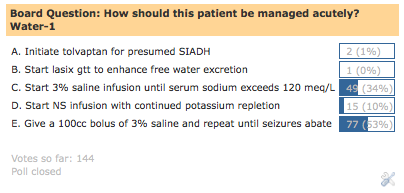
Board question of the week: Water-1

A 40-year-old woman is rushed to ER for change in mental status. She has a history of multiple sclerosis and alcohol abuse. On arrival to ER, she exhibits 3 stereotyped, tonic-clonic seizure that resolve with ativan. The initial metabolic panel is listed below:
Na- 115, K- 1.6, Cl- 56, HCO3- 40, BUN- 10, Cr- 0.8, Serum osmolality- 240 Her husband arrives at the ER and reports heavy alcohol use over the last few weeks. He brings her medication list, which includes: Protonix, Tysabri (natalizumab, anti-alpha-4-integrin antibody), and Prednisone. Blood work from 3 months prior show a normal sodium level. She received 2L of normal saline in the ER, 80 meq of KCl, and one dose of lasix. Her urine output has been 3L so far. Urine electrolytes are obtained (Una 75, Uk 10). She weighs 100kg and on examination the patient is noted to be in “status”. Nephrology is consulted for the management of her hyponatremia.

How should this patient be managed acutely?
Possible answers are listed in the poll on the upper right hand column.

The answer and explanation will be posted on Friday August 27th

Michael Lattanzio DO

*RFN board questions are meant to help introduce concepts about nephrology related diseases and do not represent actual questions seen on the ABIM exam.

Sunday, November 15, 2009

Diagnosing Reset Osmostat

The "reset osmostat" is a cause of hyponatremia, sometimes considered a variant of SIADH, in which the kidney retains its ability to appropriately concentrate and dilute the urine; however, the threshold for ADH secretion is reset downward. That is, instead of ADH being secreted with the serum osmolality increases beyond 280-285 mOsm/kg as in most individuals, it is secreted at a lower value.

Diagnosing reset osmostat is a diagnosis of exclusion. Individuals must be euvolemic, and a thorough exclusion of other causes of euvolemic hyponatremia (e.g., hypothyroidism, cortisol deficiency, medications, etc) must take place. A key feature of reset osmostat is that individuals should be able to concentrate and dilute the urine appropriately. Thus, a water challenge should result in a dilute urine (e.g., less than 100 mOsm/kg) and a water deprivation test should result in a concentrated urine. Sometimes, a patient given a diagnosis of SIADH will be proven to be reset osmostat when it becomes apparent that fluid restriction does not successfully raise the serum sodium level.

Reset osmostat classically occurs in neurologic conditions such as epilepsy and paraplegia, in addition to pregnancy, malignancy, and malnutrition. It has also been observed in healthy individuals, such as this 60 year-old man with a chronic sodium level between 125-130 mmol/L; the authors suggest that a 1951 grenade explosion the patient experienced may have caused the osmostat to reset!

Wednesday, October 7, 2009

Calculating Total Body Water

The easy way to calculate total body water is simply to multiply 0.6 times your weight in kilograms, since roughly 2/3 of your body weight is water.

There are fancier (and more accurate) equation-based calculations used to determine total body water as well.  These include the Watson formula, the Hume formula, and the Mellits-Cheek formula (specifically designed for use in children).  A handy link to a calculator using these formulas can be found here.

How were these formulas arrived at?  For the calculation of the Watson formula, for instance, dilutional methods using deuterated water (a.k.a. "heavy water") were carried out in order to obtain a "gold standard" of total body water for a large population of normal individuals; statistics were then applied to generate an equation which accurately predicts TBW based on variables such as weight, height, age, and gender.  To carry out such a study, a known dose of heavy water (D2O) is ingested by healthy volunteers and allowed to equilibrate with total body water.  Then, a mass spectrometry instrument is used to measure the deuterium:hydrogen ratio in exhaled body water vapor; the total body water can then be calculated by determining the increase in breath deuterium content in relation to the volume of heavy water taken in.  An alternative technique to determining TBW is using bioelectrical impedence analysis.  

Tuesday, September 22, 2009

Death by Soy Sauce?

Bizarre case of hypernatremia:  a suicide attempt by a 73 year-old Japanese man in which the individual drank massive amounts of soy sauce.  According to this 2006 Neurology paper by Machino et al, the patient presented with vomiting, tremor, and altered mental status, along with a serum Na of 188 mEq/L, a serum chloride of 142 mEq/L, and a serum osmolarity of 314 mOsm/kg.  MRI imaging demonstrated symmetric brain shrinkage consistent with severe, acute hyperosmolarity.  Fortunately, rapid correction of the patient's sodium (the soy sauce ingestion had apparently been within 12 hours of his initial presentation) led to rapid clinical improvement.  A similar 2004 case report by Sakai et al suggests that acute hemodialysis is another way to rapidly reverse hypernatremia as caused by acute soy sauce ingestion (over 1 Liter in this paper!). 

Friday, September 18, 2009

Aquaporins and the Kidney


The discovery of aquaporins--the protein family of water channels which regulate water transport in the kidney and many other tissues--was considered significant enough to win the Nobel Prize in Chemistry for Peter Agre in 2003.  

The aquaporins are a large family--there are at least 12 aquaporin genes in humans and possibly more--but which ones are important in the kidney?  It turns out that different aquaporins are expressed in discrete segments along the nephron.  The one which gets the most attention is likely aquaporin-2 (AQP2), which is expressed in the apical membrane and intracellular vesicles of principal cells of the collecting duct.  AQP2 is the predominant vasopressin-responsive aquaporin, and is therefore responsible for the majority of regulation of water reabsorption in the collecting duct.  According to the current model, vasopressin binds to its receptor on the basolateral surface of collecting duct cells, resulting in an increase in cyclic AMP, protein kinase A activity, and the increased trafficking of intracellular AQP2-containing vesicles to the apical membrane, where they fuse with the plasma membrane and allow water reabsorption to take place.  Mutations in the AQP2 gene account for some cases of nephrogenic diabetes insipidus. 
 
The other aquaporins also play specific roles in water transport.  For instance, aquaporins-3 and -4 are expressed at the basolateral surface of cortical collecting duct cells and are responsible for movement of water taken in by AQP2.  Aquaporin-1 is extremely abundant in the proximal convoluted tubule and descending thin limb, but is notably absent in highly water-impermeable segments such as the ascending thin limb, thick ascending limb, and distal tubule.  

Monday, June 22, 2009

Pseudohyponatremia

Hyponatremia can be broadly divided into two fundamental categories:  "true hyponatremia" (in which the serum sodium concentration is truly less than normal), or "pseudohyponatremia," in which the serum sodium concentration is actually normal but erroneously reported as low due to the presence of either hyperlipidemia or hyperproteinemia.

To understand why this is the case, it's first necessary to understand that human plasma is normally composed of 93% plasma water and 7% proteins & lipids. Furthermore, it is necessary to understand that most clinical laboratories measure sodium using an indirect ion-selective electrode (ISE) which involves diluting the original blood sample in a 1:10 ratio and measuring whole plasma sodium based on the assumption that the sample is composed of 93% water. Thus, anything which increases the protein or lipid concentration of plasma will lead to an erroneously low sodium concentration. It occurs more often with hyperlipidemia than with hyperproteinemia, and one example would be in patients with familial hypercholesterolemia where their blood is highly lipemic. 

One way to avoid measurements of pseudohyponatremia is to use a direct ion-sensitive electrode, which measures only the aqueous phase of an undiluted blood sample; however, in most labs this is not done routinely. 

Pseudohyponatremia should also be differentiated from dilutional hyponatremia, in which an osmotic shift of water from cells to the vascular space following mannitol or IVIG infusions results in true (but hypertonic) hyponatremia.

Thursday, June 11, 2009

Gestational Diabetes Insipidus

Changes in the regulation of serum osmolarity is just one of the many chances that occurs during the normal physiologic response to pregnancy.  Beginning with the first trimester, the plasma osmolarity level, serum Na, and osmolarity thresholds for the thirst and ADH responses are decreased by about 10 mosm/kg, a phenomenon which generally lasts through term.  

Later on in pregnancy, however, levels of circulating ADH actually decrease--due to production of the enzyme vasopressinase, which drastically enhances the turnover of ADH.  In most cases, the presence of vasopressinase counterbalances the decreased threshold for ADH secretion.  However, in individuals with gestational diabetes insipidus--a rare disorder in which vasopressinase production is excessive--can develop polyuria, polydipsia and excessive thirst, usually manifesting during the third trimester.  

Gestational diabetes insipidus, if severe, can be treated with DDAVP--a synthetic peptide analogue of ADH (pictured above) which is resistant to endogenous vasopressinase activity.

Wednesday, May 13, 2009

Stranded on a Lifeboat

We all know that if you are stranded on a lifeboat in the middle of the ocean, you are not supposed to drink the salt water.  Why is this the case--shouldn't the kidney be smart enough to retain the water and excrete the salt?  


It turns out that the high osmolarity of seawater (usually >1000mosm/L) either approaches or exceeds the concentrating capacity of the kidney--and therefore you can't expect to retain any free water.  In addition, seawater contains high concentration of magnesium and sulfate-containing minerals which can result in an osmotic diarrhea when large quantities are ingested; this can exacerbate free water loss which may likely already be high due to high insensible losses from wind and sun.  


Some animals have impressive adaptive mechanisms to maintain homeostasis of osmolarity in a high salt environment;for instance,  the albatross' nasal gland excretes a highly-concentrated salt solution, and the shark rectal gland has the same ability as detailed in another post.  


Plus one fun (well, not so fun for the patient involved) link for the hell of it.  The headline says its all:  Tainted Castor Oil Used for Cosmetic Augmentation Causes Kidney Failure, but you can click the link for more juicy details.

Monday, April 27, 2009

How to Do the Water Deprivation Test

The water deprivation test is useful in the workup of patients with polyuria under certain situations. The differential diagnosis of polyuria often comes down to the following: does the patient have diabetes insipidus (either central or nephrogenic), psychogenic polydipsia, or an osmotic diuresis (e.g., due to hyperglycemia for instance). The first two conditions (DI or polydipsia) are all characterized by polyuria with a dilute urine osmolarity, but how to distinguish between them? If the serum sodium is high (D.I.) or low (polydipsia) the answer may be easily apparent, but in the instance where serum sodium is within the normal range the water deprivation test can provide an answer.

However, it's important to do this test in a controlled setting, as water restriction in a patient with D.I. can result in elevated serum sodium concentrations. Patients who undergo the water deprivation test should have the urine volume and urine osmolality every hour and plasma sodium concentration eveyr two hours once water deprivation begins. The test is continued until either:

(a) the urine osmolality reaches a normal value (e.g., above 600 mosm/kg, suggesting that both ADH secretion and response to ADH are intact).
(b) the urine osmolality is stable on two successive measurements despite a rising plasma osmolality, or
(c) the plasma osmolality is greater than 295-300 mosm/kg.

In the situations of either (b) or (c), exogenous ADH is administered and the urine osmolality and volume are further monitored.

In central D.I., exogenous ADH is predicted to lead to a rapid rise in urine osmolality: in complete D.I., the urine osm will more than double, while in partial central D.I. (which is more common) there will be an increase of at least 15% in the urine osm. Generally individuals with central D.I. are able to concentrate their urine osm > 300 mosm/kg.

In nephrogenic D.I., there is either no response to ADH (complete nephrogenic D.I.) or a blunted response to ADH (up to 45%), though patients are rarely able to concentrate their urine osm above 300 mosm/kg.

A more detailed protocol for the water deprivation test, approved by the Scientific Advisory Committee of the Diabetes Insipidus Foundation, Inc, can be found here.

And for the really hard-core students of water metabolism, you can check out the following article I stumbled across on PubMed: The use of the water deprivation test for the diagnosis of apparent psychogenic polydipsia in a socially deprived African grey parrot (Psittacus erithacus erithacus), published in a 1988 issue of Avian Pathology.

Wednesday, April 22, 2009

Hypovolemia versus Dehydration

It is important to realize the difference between HYPOVOLEMIA and DEHYDRATION, two terms which are often used interchangeably but in fact reflect different things. Read the Up-To-Date entry « Dehydration is not synonymous with hypovolemia » by Burton Rose if there is any confusion about this. HYPOVOLEMIA refers to any condition in which the extracellular fluid volume is reduced, and results in decreased tissue perfusion. It can be produced by either salt and water loss (e.g. with vomiting, diarrhea, diuretics, or 3rd spacing) OR by water loss alone, which is termed DEHYDRATION. Salt + water loss comes primarily from the extracellular fluid whereas pure water loss (dehydration) come from the total body water, only about 1/3 of which is extracellular. Thus for dehydration to produce the same degree of extracellular volume depletion as salt+water loss, 2.5 times as much fluid needs to be lost. Patients with DEHYDRATION are always hypernatremic.

Friday, March 27, 2009

Survival Rule of Threes

I recently learned the "Survival Rule of Threes"--which gives a rough estimate of how long a person can live in various extreme survival conditions.  For example....

...you can expect to live for 3 minutes without air.
...you can expect to live for 3 hours without shelter (in an extreme condition like Antarctica for example).
...you can expect to live for 3 days without water.
...you can expect to live for 3 weeks without food.

Obviously these are only rough estimates (and I don't encourage you to find out whether or not you can beat these various marks) and there is a wide variability between human beings.  Gandhi was able to fast for 3 weeks while already in his 70s.  One member of the Irish Republican Army, Thomas McElwee, successfully survived for 73 days during a hunger strike.   The infamous case of Terry Schiavo in 2005 ended with her death about 2 weeks following the court-ordered removal of her feeding tube.  
Not surprisingly, one can last a lot longer without food than without water, a testament to (a) how much water one loses with just the normal processes of breathing and sweating, and (b) the potentially lethal complications of hypernatremia and the importance of maintaining appropriate water concentration.

Monday, March 23, 2009

Triphasic diabetes insipidus

In central diabetes insipidus induced by cerebral trauma or following neurosurgery, there is a "triphasic" presentation which may occur. Briefly, the patient begins with a tendency towards hypernatremia, then develops a tendency towards hyponatremia, and finally ends with a chronic tendency towards hypernatremia. The three phases are described below:


  1. First Phase: due to ischemia or direct trauma to the vasopressin-secreting neurons of the hypothalamus, there is an initial polyuric phase that lasts for about 4-5 days in which there is a fall in urine osmalality and, if the patient loses too much free water, hypernatremia ensues.
  2. Second Phase: in the second phase, there is a transient SIADH occurring as a result of leakage of vasopressin from damaged posterior pituitary tissue and severed axons. This typically occurs around days 5-6 post-event and the tendency to hyponatremia may be exacerbated by the administration of free water given in response to the First Phase.
  3. Third Phase: after all the ADH from damaged neurons has leaked out, individuals may or may not enter the third phase, a chronic diabetes insipidus. This does not happen in all individuals as over 80-90% death of all vasopressin-secreting neurons must be destroyed in order for central D.I. to occur.

Saturday, March 21, 2009

IVIG & Hyponatremia

It has been long recognized that IVIG--given for a variety of immune-mediated diseases and infections--has been associated with hyponatremia. In the recent "Electrolytes" edition of Neph-Sap, their explanation is a multifactorial one:

1. pseudohyponatremia: there is a massive protein load in an IVIG infusion (in the form of immunoglobulins), and as a result the protein phase of serum is higher. Depending on the type of laboratory test used to determine serum sodium concentration, the Na may or may not be read as low.

2. hypertonic hyponatremia: most IVIG preparations contain significant amounts of sucrose or maltose--whose presence is necessary to prevent aggregation of Ig molecules--and the resultant increase in serum osmolality will result in the efflux of water out of cells into the extracellular fluid compartment, thereby lowering serum sodium. The mechanism is similar to hyperglycemic hyponatremia or mannitol-induced hyponatremia.

3. true dilutional hyponatremia: the total volume of the IVIG infusion may be significant enough to cause dilutational hyponatremia, in large part because the high oncotic pressure allows for the solute to be retained intravascularly for prolonged periods of time.

Wednesday, March 11, 2009

Beer Potomania


One of the admittedly rare causes of hyponatremia is beer potomania--the excessive consumption of beer in the absence of much food. How does beer potomania work?

Beer has a very low content of sodium and protein--and if an individual subsists solely on beer they have a very limited solute intake. This is important because the limit on how much free water excretion can be achieved in a day is dependent on urine osmolality (urine flow rate = rate of solute excretion / urine osmolality). For an individual drinking predominantly beer, they are taking in only about 300mosm of solute per day, which is then eliminated in the urine. This puts a cap of only about 4-5 Liters of electrolyte-free water that can be excreted in a day. Thus, if an individual drinks more than 4-5 Liters of beer in a day, in the absence of additional solute, they will become hyponatremic. On top of this mechanism, beer drinkers may also have non-osmotic secretion of ADH as a result of volume depletion from chronic GI losses or vomiting. This can result in profoundly low serum sodium levels and all the neurologic complications which go along with it. The same general mechanisms (low solute intake, high fluid intake) are also at play in elderly women eating a "tea and toast" diet.

Sunday, March 1, 2009

On Gatorade


Did you know that the inventor of the popular sports drink "Gatorade" was a Nephrologist? The 2007 NY Times obituary of J. Robert Cade, the University of Florida nephrologist who concocted Gatorade, describes the story of the beginning of the now billion-dollar sports drink industry. Ironically, Dr. Cade passed away at the age of 80 of kidney failure.

As the story goes, football coaches of the Florida Gators football team were perplexed by the fact that their players were unable to urinate after football practices in the humid Florida swampy summers. Enter J. Robert Cade, who noted that this was a simple response to hypovolemia, and intended to correct the situation by having the players drink a solution of sodium, potassium, glucose and sucrose. The initial versions were substantially more salty than current versions, and as legend has it, it was not until Cade's wife came up with the idea of adding lemon to the stuff (and also, probably reducing the salt concentration) that it started to catch on. Also interesting, Cade initially offered the University of Florida the opportunity to purchase the patent rights--they initially declined, and once the sales of Gatorade began increasing, they sued Cade and there was a protracted legal battle for the rights to the sports drink.

In its current form, according to the Gatorade FAQ web page, Gatorade is stated as being slightly hyperosmolar compared to blood. However most of the osmolarity here is being provided by sugar, and once it is metabolized, it is in fact in essence a hypoosmolar solution, as Gatorade falls into FDA's category of "low-sodium" products. This is why ingestion of large quantities of Gatorade during a marathon, for instance, may be associated with the development of dangerous hyponatremia.

Oral rehydration solution--which has been attributed in decreasing worldwide death rates from diarrhea in developing countries by millions--has a much higher sodium content, 75 mmol/L according to WHO Guidelines. In contrast, Gatorade & other sports drinks have a typical NaCl concentration of between 10-25 mmol/L. In addition, the concentration of KCl is much lower in sports drinks (3-5 mmol/L) compared to that of oral rehydration solution (20 mmol/L) by WHO guidelines.

This study was a randomized controlled trial looking at the treatment of adult patients with viral gastroenteritis in the U.S. with either Gatorade, Pedialyte, or oral rehydration solution. Patients in all three groups showed similar rates of recovery, indicating that all three options are probably safe. However, the Gatorade group showed higher degrees of hypokalemia.

Here is the full composition of oral rehydration solution according to WHO, as taken from wikipedia:
Ingredient g/L Molecule mmol/L
Sodium chloride (NaCl) 2.6 Sodium 75
Glucose, anhydrous (C6H12O6) 13.5 Glucose 75
potassium chloride (KCl) 1.5 Potassium 20


Chloride 65
trisodium citrate, dihydrate Na3C6H5O7•2H2O 2.9 Citrate 10