Showing posts with label glomerular disease. Show all posts

Wednesday, August 1, 2018

Alport Syndrome


I recently saw a patient in clinic with long-standing hematuria with numerous family members on her mother’s side with hematuria.  She now presented with proteinuria but stable renal function.  Collagen IVa disease was highly suspected and genetic sequencing identified a heterozygous carrier for a previously characterized pathogenic mutation in Col4a5.

In the process of taking care of this patient who was heterozygous for X-linked Alport’s syndrome, I wondered, “Who is Dr. Alport?”.
Dr. Arthur Cecil Alport was a physician originally from South Africa who attained his medical training in Edinburgh, Scotland. He had many different interests initially studying malaria abroad and then practicing medicine in London before becoming a Professor in Egypt where he fought for the care of poor patients.  He showed that with careful observation one can provide valuable insights into a specific disease. 
Dr. Alport was not the first to identify the entity of hereditary hemorrhagic nephritis.  Initially, William Howship Dickinson described a family with 11 out of 16 members with albuminuria in 1875.  Subsequent studies by Guthrie and Hurst identified families with hematuria and kidney disease of varying severity.  Dr. Alport saw a patient from the Guthrie/Hurst cohort which he further studied and published with the title, “Hereditary Familial Congenital Haemorrhagic Nephritis”, in the British Medical Journal in 1927 which led to the identification of the disease as Alport’s syndrome. 
In this paper, he found a number of female members of the family had hematuria but did not develop edema, heart failure, and kidney failure, a fate reserved for a selected few male members of the family.  He also noted numerous female members with profound deafness that at time was not associated with hematuria.  Though he acknowledged the hereditary nature of this disease, he also found hematuria and albuminuria were exacerbated by streptococcal infection which he had limited success in recreating in rabbits. 
The history of medicine often provides an interesting context for our current understanding of human disease.  By observing the association of deafness in families with hereditary hematuria, Dr. Alport brought to light a key identifier of the disease entity.  This identification ultimately led to the disease to be associated with his name though the renal phenotype of familial hematuria was discovered by prior investigators.
Posted by Ankit Patel, Nephrology Fellow, Joint BWH/MGH Fellowship Program

Sunday, July 15, 2018

Primary Membranous Nephropathy


The landscape of Primary Membranous Nephropathy (PMN) has significantly changed over the last decade. Most of what we now understand about the pathogenesis of PMN is derived from the Heymann Nephritis model in rats. In 1959, Dr. Walter Heymann published this landmark article, which established the autoimmune nature of the disease. In the late 1970s, two groups (Dr. Couser et al in Boston and Dr. Van Damme et al in the Netherlands) established that the deposits of Heymann Nephritis were not from circulating immune complex trapping, but due to in situ formation. The rat antigen was eventually identified as megalin. The human antigen equivalent remained unknown until much later. Finally, 2009, Salant, Beck et al found that 70% of adult patients with “idiopathic” membranous nephropathy had antibodies against M-type phospholipase A2 receptor (PLA2R), a transmembrane glycoprotein found on podocytes. Soon after, another podocyte transmembrane glycoprotein called thrombospondin type-1 domain-containing 7A (THSD7A) was discovered (See NEJM 2014; 371(24):2277-2287).

Now, approximately 85% of PMN is found to be mediated by anti-PLA2R or anti-THSD7A, with still 15% of PMN “idiopathic”. With a serologic test for anti-PLA2R now commercially available as well as a biopsy immunofluorescent stain, the diagnosis and management of patients with PMN has been completely revamped.

For more on this discussion, please join us in the next GlomCon Interactive Fellows’ Curriculum on Tuesday, July 17th at 11 am eastern time. We will be walking through a case scenario as we review several different aspects of PMN.

To join the meeting go to: https://glomcon.zoom.us/ and select 'Join a Meeting'. Enter the Meeting ID into the web site (or connect directly from your Zoom app).
Meeting ID: 394-801-817
You HAVE to be logged into ZOOM to join the session. You may either create a free ZOOM account, or sign in through a Google or Facebook account (if you have one).   

Alternatively, use your institution's 'Polycom' system, enter IP address:
US West: 162.255.37.11
US East: 162.255.36.11
Europe: 213.19.144.110
Then, enter the Meeting ID: 394-801-817

Monday, June 11, 2018

Minimal Change Disease


The next session of GlomCon's Nephropathology Essentials will be held this Tuesday, June 12th at 11 am EDT. Dr. Helmut Rennke will be leading the session which will focus on Minimal Change Disease and FSGS (see below for session details).

Minimal change disease (MCD) is the most common cause of nephrotic syndrome in children > 1 year of age. Adult MCD is much less common but remains an important cause of nephrotic syndrome in this population. Here are some quick points to remember in adults with MCD:
  • Bimodal presentation: young children and very old adults
  • The slight male predominance (2:1) found in children disappears in the adult population
  • More common in atopic patients
  • Clinical presentation: Abrupt onset of edema, nephrotic range proteinuria, severe hypoalbuminemia and severe hyperlipidemia
  • There is a long list of secondary causes of MCD, but important ones to remember include: NSAIDs, Lithium, and Lymphoma 
  • Microscopic hematuria is seen in 10-30% of cases
  • AKI is present in 20% of cases (higher risk in older age, males, and presence of HTN)
  • Best initial treatment is corticosteroids 
    • Prednisone 1 mg/kg (maximum 80 mg) daily or alternative-day dosing of 2 mg/kg (max 120 mg)
  • Time to complete remission is much longer in adults, requiring 12-16 weeks
  • Relapse rates in adults with MCD are high
This is reviewed nicely in the following articles

MCD was also covered in this past GlomCon session. For further on both MCS and FSGS pathology, make sure to join the session on Tuesday.

GlomCon's Nephropathology Essentials Details:
To join the meeting go to: https://glomcon.zoom.us/ and select 'Join a Meeting'. Enter the Meeting ID into the web site (or connect directly from your Zoom app).
Meeting ID: 394-801-817
You HAVE to be logged into ZOOM to join the session. You may either create a free ZOOM account, or sign in through a Google or Facebook account (if you have one).  
Alternatively, use your institution's 'Polycom' system, enter IP address:
US West: 162.255.37.11
US East: 162.255.36.11
Europe: 213.19.144.110
Then, enter the Meeting ID: 394-801-817

Posted by: Pravir Baxi, Rush University Medical Center

Sunday, June 3, 2018

GlomCon Nephropathology Essentials


The goal of the GlomCon Teaching Platform is to enable Nephrologists, Nephropathologists, and trainees in Medicine, Nephrology, and Pathology to connect with each other to 1) Discuss challenging cases, 2) Share clinical experiences and 3) Obtain the expertise of participating faculty and invited speakers. Access to this resource is completely free!

Going forward, every Tuesday (same time weekly, 11am EDT) there will be a case-based conference, fellow's interactive conference, or renal pathology seminar.

You are invited to the inaugural session of GlomCon Nephropathology Essentials on Tuesday, June 5th at 11 am EDT. The first session will be led by Dr. Isaac E. Stillman and will focus on the basic principles of renal pathology.

For more on the Nephropathology Essentials webinars, visit: https://glomcon.org/nephropathology-essentials/

To join the meeting go to: https://glomcon.zoom.us/ and select 'Join a Meeting'. Enter the Meeting ID into the web site (or connect directly from your Zoom app).
Meeting ID: 394-801-817

You HAVE to be logged into ZOOM to join the session. You may either create a free ZOOM account, or sign in through a Google or Facebook account (if you have one). 

Alternatively, use your institution's 'Polycom' system, enter IP address:
US West: 162.255.37.11
US East: 162.255.36.11
Europe: 213.19.144.110
Then, enter the Meeting ID: 394-801-817


Monday, June 23, 2014

IgM Nephropathy: What You Need to Know


Similar to my recent post on C1q Nephropathy, IgM Nephropathy (IgMN) is a podocytopathy that is often considered a variant of Minimal Change disease (MCD) or FSGS. Like C1QN, it is also a controversial entity with a lack of uniformity from a diagnostic point of view. This undoubtedly leads to under-reporting with some pathologists calling a case IgMN what others would call MCD/FSGS/Mesangial Proflierative GN with low level IgM staining.
First descriptions of IgMN reported mesangial hypercellularity on light microscopy, granular IgM and C3 mesangial deposits on immunofluorescence with electron dense deposits in about half of cases. Podocyte foot process effacement was also evident consistent with the presentation of nephrotic syndrome. The incidence has been reported to be 2-18% of renal biopsy series and age at presentation appears to be bimodal with a peak in childhood and again later in life, 6-7th decades. The incidence of IgMN may be higher in the developing world and most recent reports come from developing nations (ref, ref).
The accurate diagnosis of IgMN is important from a prognostic and therapeutic point of view. Overall, IgMN does not respond to steroids as well as MCD with a higher proportion of patient with steroid-dependent and resistant disease. Furthermore, assessment of children with steroid-dependent and resistant MCD revealed a high proportion of IgMN, which appeared to respond better to cyclosporin than to cyclophosphamide. Rituximab (as ever!) may be another alternative therapeutic choice, although evidence remains at case report level, including a case of recurrent IgMN post-transplant. Similar to C1QN, the severity is likely dependent on the light microscopic changes with MCD patterns reporting an excellent remission rate and an FSGS pattern of injury having a worse prognosis (ref). Most cases however may have mesangial proliferative changes evident. There is even a report of IgMN presenting with crescentic lesions.

IgM deposition may be seen in many other glomerular disorders and its role is unclear. Is it an innocent bystander, reflecting natural IgM binding to exposed epitopes on injured glomerular cells? There is some evidence that IgM deposition may activate complement and that efforts to prevent IgM antibody deposition may prevent complement activation and slow the progression of glomerular injury. IgMN is another glomerular disease that we have an unclear understanding of pathogenesis and etiology where we rely on crude pathological descriptions. Hopefully the future will bring clarity to these diseases and facilitate precise molecular diagnosis of entities such as IgMN.

Saturday, June 14, 2014

C1q Nephropathy: What You Need to Know

I saw a 20 year old African American male in clinic with nephrotic range proteinuria, reduced GFR and no signs or symptoms of a systemic disease. His mother developed ESRD at age 36 and his brother, aged 26, has CKD Stage 4. A full laboratory workup including complement proteins and ANA was unremarkable. Renal biopsy demonstrated an FSGS pattern of injury with a full house picture on immunofluorescence but with dominant C1q staining. A diagnosis of C1q Nephropathy (C1QN) was made. 

Jennette et al described C1QN in 1985 in 15 patients without evidence of lupus who had dominant mesangial C1q deposition, often with C3 and Ig’s also. The mean age at presentation was almost 18 years, with a similar male: female ratio and average proteinuria of 7.5g/day. Another series from Columbia University included 19 patients who were predominantly African American (73%), young and female (73%) with nephrotic range proteinuria. Renal outcomes across the series’ (including this European series) are mixed but would suggest immunosuppressive treatment directed against the primary pathology should be considered.
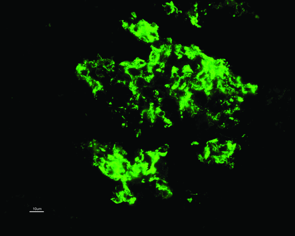
C1QN may be considered an independent pathological entity, although this has been controversial, with some classing it as a subtype of FSGS or minimal change disease (MCD). The prevalence varies (0.2-16%) but is more common in children. The predominant light microscopic appearance may be MCD, FSGS or a proliferative glomerulonephritis. While C1q deposition (predominantly in the mesangium; see image) is dominant or co-dominant by definition, a full house pattern of IgM, IgG, IgA, C1q and C3 may be observed, more commonly with a proliferative glomerulonephritis. As for treatment, the case series are small and largely uninformative. Some patients appear to remit with or without immunosuppressive treatment whereas others have a rapid course towards ESRD. Patients with an FSGS pattern may do worse than other types. For what it’s worth, my patient had no response to 6 months of steroids and mycophenolate and a plan for cyclosporine was made. The fact he likely has a genetic element to his disease adds complexity. Some would argue that immunosuppressive therapy in these cases is not likely to be of benefit but we see from our hereditary FSGS cohort that this is not necessarily the case. Cyclosporine and Rituximab in particular appear to have a directly beneficial effect on podocyte function (also see case report of Rituximab treatment in C1QN).
As for the pathogenesis and significance of the C1q deposits, we have much to learn. Are the C1q deposits pathogenic or merely markers of injury? We do know that C1q binds to the basement membrane via laminin and C1q receptors enhance binding of immune complexes to mesangial cells where it combines with other complement proteins to form C1 protease. This in theory allows activation of the classical complement cascade. Despite this, serum C4 levels are generally normal and I can find no reports of complement directed therapy (Eculizumab or C1-Inhibitors) in C1QN.
Bottom line: C1QN is likely an under recognized entity which can have varying clinical and histological presentations. Renal outcomes are also variable and it may have a different prognosis compared to traditional MCD (& FSGS?). One to keep in mind for the boards.

Sunday, November 17, 2013

Abatacept for Glomerular Diseases: A New Era of Intelligent immunosuppression?

At ASN Kidney Week there was some interest in abatacept as a targeted therapy for glomerular diseases. T-cell activation requires 2 signals; (i) binding of the T-cell receptor to the antigen-MHC complex on the antigen-presenting cell and (ii) a co-stimulatory signal involving CTLA-4 on the T-cell and B7-1 on the antigen presenting cell. Abatacept is a fusion protein composed of the Fc region of IgG1 fused to the extracellular domain of CTLA-4 which inhibits the T-cell co-stimulatory pathway via B7-1 binding. 

          FSGS
The headlines must go to the small case series of abatacept in FSGS published in NEJM. The rationale for its use was the observation that B7-1 expression is not apparent in normal human podocytes but is found in certain diseased podocytes including a subset of FSGS patients. The series included 4 patients with recurrent FSGS post-transplantation (rituximab-resistant) and one with glucocorticoid-resistant primary FSGS. All patients achieved either partial or complete remission.
In vitro studies demonstrated that α3-Integrin knockout mice constitutively expressed B7-1 in podocytes and abatacept blocked B7-1 mediated podocyte migration in these cells. The molecular mechanism of B7-1-induced podocyte dysfunction was shown to be disruption of activation of the glomerular protein β1-integrin. The authors conclude that B7-1 immunostaining of biopsies may identify a subgroup of patients who would benefit from treatment with abatacept.

2       Lupus Nephritis
The late-breaking session included a randomized controlled trial of Euro-lupus regime cyclophosphamide (i.e. low dose IV) with or without abatacept for proliferative lupus nephritis [Access Trial]. Azathioprine was introduced at 3 months and stopped at 6 months in the abatacept group if they had achieved a remission. Overall, there was no difference in remission rate between the groups. Despite the neutral outcome, 2 points should be taken from the study: (i) The Euro-lupus regime appeared to work in a US cohort of patients where almost 80% were either Hispanic or African American. (ii) Abatacept patients who achieved remission maintained this at 1 year despite coming off immunosuppression at 6 months. However, with the growing confidence in Mycophenolate-based therapy for lupus nephritis and the lack of improved remission with Abatacept in this study, its place in the treatment of proliferative lupus nephritis remains uncertain.

      Diabetic Nephropathy
An oral presentation on abatacept in Diabetic Nephropathy [FR-OR010] reported increased B7-1 expression in both murine podocytes cultured in high-glucose and on human glomerular podocytes from biopsy specimens. The use of Abatacept in diabetic mice prevented an increase in albuminuria.

Bottom Line: The FSGS case series beautifully illustrates how targeted therapies may be applied to immune-mediated renal diseases. While this case series is very small, it demonstrates the potential for reclassifying disease based on pathogenesis (i.e. B7-1-mediated) rather than crude pathological patterns (focal segmental sclerosis). This is similar to the recent re-classification of MPGN into complement or immune complex-mediated forms. With new targeted therapies like abatacept (and eculizumab for complement mediated glomerulopathies), we may be entering an era of intelligent immunosuppression based on molecular pathogenic signals rather than crude histological patterns.

Sunday, September 15, 2013

Genetic Defects of the Glomerular Basement Membrane

The glomerular basement membrane is a thin layer of extracellular membrane proteins that is an important part of the filtration barrier, particularly glomerular permselectivity, by preventing proteins from crossing into the filtrate. The major proteins in the GBM are laminin, type IV collagen, nidogen and the heparan sulphate proteoglycan agrin. Interestingly, in the past, I was taught that the highly negative charge on agrin played the major role in mediating charge selectivity. However, recent studies have shown that mutations in the gene encoding agrin, leading to a reduction in charge along the GBM, have no effect of glomerular function in mice. Similarly, deletion of agrin has little effect on permselectivity further suggesting that the role of the proteoglycans in selective filtration is minor at best.

There are two conditions associated with genetic defects in glomerular basement membrane proteins:


Syndrome
Gene(s) affected
Protein
Phenotype
Alports Syndrome
COL4A3
COL4A4
COL4A5
Type IV Collagen, α3, α4, α5 subunits
Initial normal formation of GBM but eventually hematuria, proteinuria and eventual ESRD with characteristic splitting of the GBM. COL4A5 mutations are commonest and are X-linked. Other forms are autosomal.
Pierson Syndrome
LAMB2
Laminin β2
Autosomal recessive disorder with variable phenotype depending on the particular mutation. However, ocular abnormalities (microcoria) are present at birth and the majority of affected individuals progress to ESRD within the first few weeks/months of life. Extrarenal manifestations including hypotonia and neurodevelopmental defects have been reported.


Please see this excellent review in Nature Reviews Nephrology for further information.


Friday, June 28, 2013

Lonely Glomeruli

One of the difficulties in doing molecular research on the kidney is the diversity highly specialized cells that exist in the glomerulus. As a result, it is important to be able to isolate glomerular tissue from the surrounding kidney. A recent paper in KI detailing a method for isolated podocytes reminded me of a relatively simple technique that I was taught a few years ago for glomerular isolation in mice.

The technique was first described in this paper from 2002 but in brief, it involves injecting the mouse heart with deactivated magnetic beads (after euthanizing them of course). Some of these beads (which are just 5µm in diameter) get trapped in the glomerular capillaries. The kidneys are then removed, minced, digested and passed through a 100µm strainer to remove any larger particulate matter. Finally, the remaining tissue is suspended and exposed to a magnet to pull the glomeruli (with the beads inside) out of the mixture. The glomeruli are then left stuck to the wall of the tube next to the magnet and they can be easily removed.

The picture below is a low-power view of the glomeruli following isolation. You can see that there is very little non-glomerular tissue present, which is remarkable given that the glomeruli make up such a small proportion of renal tissue.


Below is a higher power view of 3 more glomeruli following isolation. You can clearly see the microbeads trapped in the glomerular capillaries. Cool science. (Click on any image to enlarge)