Showing posts with label ICU Nephrology. Show all posts

Thursday, March 2, 2017

Dialyzing a patient with an intracranial hemorrhage

Patients with intracranial hemorrhages often develop cerebral edema, which can create unique challenges when providing maintenance hemodialysis. Conall also covering this on RFN a few years back. Hemodialysis can worsen cerebral edema through a rapid decrease in serum osmolarity (urea is rapidly cleared from the blood by the dialyzer but urea transport across brain cell membranes lags behind). As a result, a fairly rapid increase in brain water content develops. Besides worsening cerebral edema, maintenance hemodialysis can also reduce cerebral perfusion if systemic hypotension occurs during dialysis. For these reasons, patients either require continuous renal replacement therapy or modified hemodialysis as outlined below. To reduce the risk of worsening cerebral edema, hemodialysis should be modified to include
  • small dialyzers
  • slower blood flow rates
  • slower dialysate flow rates
  • reduced dialysis times
To further reduce changes in osmolarity, one should
Other details to reduce overall risk include
  • cooling dialysate temperatures to reduce risk of hypotension
  • avoiding heparin administration. 
As an aside, patients with intracranial hemorrhage are often treated with hypertonic saline. Dialysis patients, in particular, are at risk of developing intravascular volume overload as a result of the large sodium load. This can lead to not only peripheral edema but also pulmonary edema, making ventilation difficult. An alternative to hypertonic saline administration or as an adjunct to reduce the overall sodium load is hyperventilation without humidified air. This will slowly create the desired hypernatremia from free water loss. Something to consider the next time you see a hemodialysis patient with an intracranial hemorrhage.

Melissa Makar, Nephrology Fellow, Duke

Tuesday, October 1, 2013

CKD after AKI in the ICU

I give a regular talk to the residents in the ICU on CRRT and one of the things that I focus on is prognosis. We all know that the outcomes of patients requiring CRRT in the ICU are poor. Multiple studies have shown that the mortality is 40-60% and that this mortality rate has not changed in the last 20 years. However, something that residents are less aware of is that, in the event that a patient survives their stay in the ICU, the majority will not require long term dialysis - approximately 80%. This is sometimes difficult to appreciate when you see patients on HD at discharge from the ICU but most of these will recover at least some renal function. One question, however, is how much function they recover and if this has any bearing on their overall mortality.

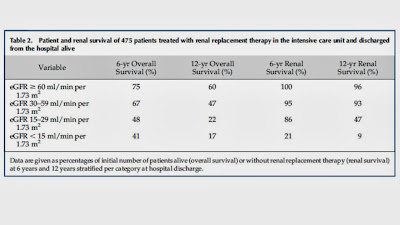
A paper recently published in CJASN goes a long way towards answering these questions. This was a retrospective cohort study of all 1220 patients admitted to the ICU requiring CRRT in a single center in the Netherlands between 1994 and 2010. As expected, the in-hospital mortality was high (55%). Of those who survived, 12% did not recover enough renal function to come off dialysis after discharge.

The commonest reasons for admission were thoracic surgery and sepsis. 20% of patients had pre-existing CKD, 48% had normal baseline renal function. There was no baseline in the remainder. At the time of discharge from hospital, 60% of patients had some degree of renal dysfunction (30% eGFR 30-60, 15% eGFR 15-30, 15% eGFR 0-15 including the 12% on HD). Of note, more than half of the patients with an eGFR <15 at discharge had pre-existing CKD. Unadjusted patient and renal survival is shown in the table:


The independent predictors of long term mortality were age, a surgical diagnosis, malignancy and an eGFR < 30. Similarly, the predictors of future need for dialysis were pre-existing CKD, and an eGFR < 30 at discharge. Interestingly, an eGFR between 30 and 60 was not associated with an increased risk of mortality or need for RRT in the future, relative to those with normal renal function at discharge.

This study adds to our knowledge of the predictors of outcomes after an episode of AKI requiring CRRT. No-one should be surprised that patients with significantly reduced GFR at discharge at are increased risk of mortality and need for eventual dialysis. However, it is reassuring that, in those patients who have an eGFR >60 at discharge, the likelihood of them requiring dialysis in the future is very low. It would be interesting to know if the presence of proteinuria modified the relationship between eGFR and mortality/need for dialysis, particularly in those with an eGFR between 30 and 60 at discharge but unfortunately, these data were not available.

Sunday, July 21, 2013

Hypothermia Protocol and Dialysis


I recently received an inpatient consultation to see a CKD 5D patient. The reason for consult, as is mostly the case with dialysis patients was that he “needs hemodialysis”.
This dialysis patient wasn’t the average bear though. He had had a witnessed cardiac arrest, was treated by EMS, and defibrillated. He had a return of spontaneous circulation after being pulseless for 20 minutes. As soon as he got to the ER, he was initiated on our standard institutional therapeutic hypothermia protocol.  I was called in to dialyze him because (it wasn’t his usual day) the cardiologist wanted to perform a left heart cath on him the following day, and they “did not want dialysis to interfere with that schedule”. My clinical assessment did not reveal a severe degree of volume overload. He wasn’t hyperkalemic, and had only a mild degree of lactic acidosis that was nicely compensated by him being appropriately ventilated. Due to the concerns that I talk about below, I did not see an emergent reason to dialyze him.
I would like to focus on a few teaching points from a nephrologist’s perspective that I took away from this scenario:
  1. Therapeutic hypothermia entails cooling post cardiac arrest patients to 32-34 degrees Celsius, ideally within 6 hours of a cardiac arrest.  Both intravascular and surface cooling methods are used. At my institution, the protocol involves administering up to 3 liters of 0.9% saline (which has been cooled to a temperature of 4 degrees Celsius), over an hour. This is complemented by cooling vests. Once target temperature is reached, the cooling phase is continued for 12-24 hours, after which the patient is rewarmed gradually at the rate of 0.5 degrees Celsius/hour.
  2. Sub-physiological body temperatures expectedly have adverse effects. Hypothermia can hamper leukocyte function, increasing infection risk later. Cardiac effects include bradycardia and prolonged QT interval (both were present in this patient). Finally, for us nephrologists, here are some adverse effects and pertinent points that we need to keep in mind for such patients:
  3. Hypothermia can cause hypokalemia via two different mechanisms. Low temperature causes a transcellular shift of potassium in to the intracellular compartment. This effect is possibly mediated by increased beta adrenergic and sympathetic activity. In fact, hypokalemia in the setting of hypothermia must be repleted extremely cautiously, if at all, given the risk of rebound hyperkalemia as potassium moves back out of the cells when the patient is rewarmed. This rebound hyperkalemia can be frequently fatal due to arrhythmias.
  4. The second mechanism by which hypothermia causes hypokalemia is by the induction of polyuria, also known as “cold diuresis”. This hypokalemia is mediated by increased urinary flow, and is seen in conjunction with hypovolemia, hypophosphatemia, and hypomagnesemia. I didn’t observe any of these in my patient, maybe because of his oligo-anuric status at baseline. Nevertheless, close monitoring of volume status and electrolytes is required.
  5. Hypothermia interferes with platelet function and with the clotting cascade. In fact, as per this review, 22% of patients had bleeding post-hypothermia induction. That might be a concern when making the decision to dialyze post-hypothermia patients with heparin.
  6. The other issue that I ran in to, that was specific to dialysis patients, was the concern about the patient’s temperature. As we know, most HD machines warm blood before returning in to the patient. With most machines, the warmer cannot actually be turned off and only goes as low as 35 degrees Celsius. In other words, dialysis can inadvertently warm the patient up to this temperature (from the target temp of 32 degrees, per the hypothermia protocol)! CRRT machines do have adjustable temp settings that goes down to 32 degrees, so that might be a safer alternative. Given the risk of inadvertently warming the patient, and because I did not see any emergent indication for dialysis, I did not dialyze the patient. I believed that in that situation, his hypothermia protocol took precedence over dialysis.
In my experience, I have observed that referring non-renal physicians often consider inpatient hemodialysis an ancillary service, akin to placing an order for an x-ray or a lab draw. Seasoned fellows have heard this phrase all too often, “I want you to come down and dialyze this patient”. You are then left with the unenviable task of explaining to the non-renal physician that the decision to dialyze would be made by the nephrologist after proper assessment of the patient (isn’t why they consulted you in the first place?). Let’s not allow our familiarity and comfort with dialysis technology lull us in to putting our guard down. Dialysis is an inherently intense and complicated procedure where multiple clinical parameters need to be closely watched. It’s a fact that is often lost in translation.
Posted by Veeraish Chauhan

Sunday, July 7, 2013

Renal Grand Rounds - Case of the month


A young woman with h/o polysubstance abuse and seizures was admitted to hospital with status epilepticus. She was treated with a propofol infusion at 5mg/kg/hr which was maintained due to perceived continuing seizure activity. On hospital day 5, she developed an acute metabolic deterioration – rhabdomyolysis : CPK 100,000, AG metabolic acidosis (HCO3 18) and non-oliguric AKI. Her EKG also became abnormal (RBBB). The diagnosis was propofol infusion syndrome.

What is propofol Infusion Syndrome (PRIS)? 
Like many syndromes, PRIS is a conglomeration of clinical and biochemical manifestations.  Based on  83 case reports from 1992-2007, PRIS is characterized by:
1) Metabolic acidosis, (pH less than 7.30 or HCO3 less than 19)
2) Rhabdomyolysis (CPK more than 10K)
3) Renal failure
4) Cardiac dysfunction (Brugada-like EKG pattern, asystole, PEA, sustained VTs, heart failure, or bradycardia) 
5) Hypertriglyceridemia (TG more than 400)
6) Hyperkalemia
7) Hypotension (or use of vasopressor agent)
8) Hepatic transaminitis
9) Hypoxia (PO2 less than 60mmHg). 

The first 5 manifestations are the most common. While there is no requirement for the number of clinical manifestations a patient must exhibit to meet diagnosis for PRIS, a prospective study showed that most patients exhibit at least 3 defining manifestations within 3 days of propofol use.  In the same study, the incidence of PRIS was found to be a low 1% --similar to other estimates.  However, PRIS is associated with high mortality, up to 30% in some studies. Moreover, because many of these manifestations are common, the presence of any of them could be attributed to another etiology, thus delaying diagnosis of PRIS.  For instance, in the case vignette, the initial rise of CK was attributed to seizure rather than PRIS.

Pathophysiology of PRIS
Inhibition of electron flow along the mitochondrial electron transport chain/ impairs oxygen utilization. Propofol or its metabolites inhibits fatty-acid oxidation leading to buildup of toxic fatty acid intermediates. As described in this nice review.

Risks of PRIS?
Critical illness (especially, CNS illness); Use of propofol dosage more than 4 mg/kg/hr —the usual adult maintenance dose is 0.3 to 3mg/kg/hr; duration of Propofol use greater than 24hr; exogenous cathecholamines and corticosteroids; poor intake of carbohydrate, see this reference.

Management of PRIS
Early recognition/diagnosis; Cessation of propofol infusion; Cardiopulmonary support; Hemodialysis (strongly advocated by expert opinion and outcome of case series).  However, there are no known RCT of use of renal replacement therapy in the treatment of PRIS.  Nonetheless, in case series, survivors of PRIS are more likely to have received HD/CVVH.  Therefore, prolonged use of HD/CVVH is worth considering by renal consult service.  Because propofol is lipophilic and has volume of distribution of 20-40L, it is poorly cleared by HD/CVVH, prolonged RRT is likely needed for PRIS management.

The patient in the case was treated with CVVH overnight, and then transitioned to intermittent HD.  At the time of her discharge, she was off HD with her Cr back to baseline.

See this previous post by Nate on PRIS.

Posted by Opeyemi Olabisi

Tuesday, February 26, 2013

Πάντα ρεί - once more on the (right) fluids


Yet another manuscript evaluating fluids for IV replacement was published in JAMA this week underscoring the importance and controversial nature of the topic. A large meta-analysis involving 10,868 patients from 38 trials showed that hydroxyethyl starch (HES) was associated with an increased risk of mortality and renal failure (RR 1.27; 95% CI 1.09 - 1.47). Of note, a recent large trial published in the NEJM in 2012 did not show an increased mortality but more patients who received resuscitation with HES were treated with renal-replacement therapy.

HES is mainly used by anesthesiologists and surgeons and has the potential advantage of decreasing the amount of total administered volume and sustaining intravascular volume for longer periods of time. The discussion about mortality and renal failure associated with HES has been going on for a long time, as has the discussion about the relative benefits of colloids vs. crystalloids. Despite the lack of strong evidence of superiority of HES over crystalloids, the clinical use has been increasing - even with the associated higher cost and safety concerns.

A major drawback for the supporters of HES was the realization that one of the leading authorities in the field and major proponent of HES, Joachim Boldt, has conducted one of the biggest cases of fraud in anesthesia with “false data published in at least 10 of the 91 articles examined, including, for instance, data on patient numbers/ study groups as well as data on the timing of measurements“. 80 articles have been retracted because the research was deemed unethical. A Cochrane review from 2012 did not find a significant difference in mortality even when the fraudulent studies were excluded. However, the current meta-analysis not only excluded the studies by Boldt, but also included 3 trials published in 2012 contributing more than half of the patients to the collective examined, thereby adding more weight to their analysis over prior ones. There was no difference in mortality when the Boldt papers were included in the analysis.

Side effects of HES mainly occur in the kidney but the exact pathophysiology of AKI associated with HES is unclear. Vacuolization as an injury pattern can be observed as discussed in an earlier post in this blog. In vitro HES showed a dose-dependent decreased viability of HK-2 cells (human immortalized proximal tubular cells) after incubation with HES130/0.4. A necropsy study suggested HES accumulation in the kidney might cause toxicity. 

What can we learn from this? Rigorous analysis and ongoing discussion and evaluation of data are of crucial importance in Medicine and scientific fraud, driven by motives such as recognition and money can cause data to shift to the wrong direction. There does not seem to be a major advantage of using HES over crystalloids and data on mortality and renal failure are tied between no difference and increased mortality/AKI in the HES group. From my perspective there seems to be no indication to use HES and this might be better for the kidney.

Posted by Florian Toegel

Monday, November 12, 2012

Spare the Chloride


Fluid therapy is essential in ICUs and not surprisingly there is still much controversy about which fluid to use, how much and when. Nephrologists often roll their eyes at other subspecialty's preferences, e.g. surgeon's preferences for Ringers, citing the risk of hyperkalemia in renal failure patients given Ringers. I learned that normal saline is the preferred agent unless there is a special consideration such as acidemia necessitating alternatives. Now chloride, the partner of sodium that gets considerably less attention most of the time, enters the stage.
Yunos et al in JAMA suggest that too much of chloride increases acute kidney injury (AKI) episodes in tertiary ICUs and increases the need for renal replacement therapy (RRT) but does not affect mortality.
The physiological rationale for the detrimental effect of chloride on the kidney is described as vasoconstriction mediated by chloride in dog experiments and a possible role of tubuloglomerular feedback mediated vasoconstriction as well as decrease in GFR caused by increased distal chloride delivery. Furthermore they cite thromboxane mediated vasoconstriction caused by chloride and enhanced responsiveness to vasoconstrictor agents as possible physiological sequelae of chloride administration.
The authors of the JAMA article conducted a prospective, open-label sequential pilot study of patients admitted consecutively to the ICU. Initially patients were treated with chloride-rich IV fluids (0.9% saline, 4% succinylated gelatin solution or 4% albumin solution) and after that initial control period a chloride-restricted strategy was implemented with lactate (Hartmann solution), a balanced solution (Plasma-lyte 148) or chloride-poor 20% albumin as preferred agents.
The results were a lower increase in serum creatinine levels and fewer episodes of RRT in the chloride-restricted group but no differences in mortality, hospital or ICU length of stay or need for RRT after discharge.
How does this study affect our choice of ICU fluids? Certainly, these results are hypothesis generating and important but need to be viewed as preliminary given the design of the study. An accompanying editorial by Waikar mentions the Hawthorne effect as potential major concern. Clearly these important preliminary data need follow up in a controlled prospective trial. 
Posted by Florian Toegel