Saturday, February 28, 2009

Nephrology Nobel Prizes

The Nobel Prize was first awarded in 1895. A number of awards have been given to individuals whose research is inseparable from the field of Nephrology. Here are a few of the notable examples:

1. 2003 Nobel Prize in Chemistry: Roderick Mackinnon and Peter Agre, for their work on ion channels and the discovery of water channels (the aquaporins) specifically. Prior to the discovery of aquaporins, most scientists believed that water merely "leaked" through the cell membrane. The highly regulated process of water transport in the collecting duct of the nephron was one important clue that specific channels are responsible for the movement of water in and out of the cell.

2. 1990 Nobel Prize in Physiology & Medicine: Joseph Murray, the surgeon who performed the first-ever kidney transplant between identical twins, thereby demonstrating that previous failures at kidney transplantation were due to immunologic incompatibilities rather than surgical technique.

3. 1997 Nobel Prize in Chemistry: Jens Skou, a Danish chemist who is credited with the discovery of the Na/K ATPase. Although this has broad implications for all cell types, the Na/K pumps is essential in understanding the chemical gradients which allow for ion transport to occur all along the nephron.

Obviously there are a ton of other Nobel prize recipients whose work has relevance to Nephrology, as well as just about every other field of medicine (e.g., the discovery of DNA by Watson & Crick) but I've listed the ones that stand out to me as being of special interest to nephrologists.

Friday, February 27, 2009

Renal (Cerebral) Salt Wasting versus SIADH

The term "cerebral salt wasting" has always been a confusing one to me, and in the most recent issue of C-JASN, the authors of an article "More on Renal Salt Wasting Without Cerebral Disease : Response to Saline Infusion", the authors advocate abandoning the term altogether in favor of the term "renal salt wasting." They make this suggestion based on the identification of a subset of patients with salt wasting in the absence of cerebral disease.

Whether renal salt wasting (RSW) even exists has been the matter of controversy. In this paper, the authors compare and contrast a patient with SIADH with another with RSW. The distinction is a tricky one to make, but important as the treatment for SIADH is water restriction whereas the treatment for RSW is saline infusion.

SIADH, as we all know, is due to the inappropriate secretion of ADH which results in unregulated water absorption; patients are generally euvolemic on clinical examination. Usually (but not always) the urine Na is >20 mmol/L and there is an elevated fractional excretion of urate (FE-urate) on initial presentation. Once the sodium corrects, however, the FE-urate normalizes as well.

RSW, on the other hand, is due to a defect in tubular sodium transport that results in sodium wasting and resultant hypovolemia. The serum renin and aldosterone levels at presentation are elevated. Urine Na is >20 mmol/L because the tubular loss of Na is the primary cause of extracellular volume depletion. The entity is differentiated from garden-variety pre-renal azotemia based on the presence of other abnormalities indicative of other defects in renal tubular function: for instance, RSW patients commonly have an elevated FE-phosphate (>20%) and elevated FE-urate whereas patients with pre-renal azotemia tend to have reduced FE-phosphate & urate.

The authors claim that it is possible to differentiate between SIADH and RSW based on the response to correction of the serum sodium: in SIADH, the FE-urate should normalize, whereas in RSW, the FE-urate remains elevated. I understand the logic, but what would be more helpful would be to differentiate between the two entities during the initial patient encounter. Perhaps FE-phosphate is one way to do this--though I'm not sure if the test has a quick enough turnaround time to be useful in this way. Assessment of volume status is another possibility, though difficult even for seasoned clinicians in many instances.

Thursday, February 26, 2009

Interesting Case of Adult Henoch-Schonlein Purpura

Saw a case in the clinic today of a 40-ish year old woman who was diagnosed about a year and a half ago with Henoch-Schonlein purpura. This is notable in that usually this is a pediatric disease--though as this case illustrates it can certainly affect adults. This particular patient had all three elements of the "classic triad" of HSP: purpuric rash, abdominal pain, and arthritis. In addition, she also experienced renal failure secondary to glomerulonephritis, with a creatinine that had elevated to 2.5mg/dL from a normal baseline and whopping proteinuria with a urine protein:creatinine ratio of >20. Fortunately, with plasma exchange, prednisone, and Cytoxan, her symptoms eventually resolved, and today in the clinic (about 1.5 years after having been initially diagnosed) her creatinine was down to 1.2 mg/dL. The disease appears to lie along the same spectrum as IgA Nephropathy, as renal biopsy of HSP patients is often indistinguishable from that of IgA Nephropathy kidneys: mesangial proliferation with IgA deposits. HSP is best categorized as a vasculitis and it is commonly preceded by an upper respiratory tract infection--it has been linked to Strep infection, viral infection, and even certain medications, though in most cases no underlying etiology causing HSP is identified.

Wednesday, February 25, 2009

Churg-Strauss Syndrome

Churg-Strauss Syndrome lies within the spectrum of Wegener's Granulomatosis, microscopic polyangiitis, and ANCA-associated vasculitis: all diseases which result from inflammation of small-to-medium-sized vessels. The "classic triad" of Churg-Strauss syndrome is asthma, eosinophilia, and vasculitis, and although "renal involvement" is not amongst these, it does occur in a subset of Churg-Strauss patients.

In this 2006 AJKD article looking at 116 Churg-Strauss patients, about 25% had some type of renal involvement. Of those who had rapidly progressive glomerulonephritis and crescents on biopsy, all were ANCA-positive. Renal involvement in Churg-Strauss should therefore generally be managed as an ANCA-associated vasculitis, e.g. steroids, Cytoxan, with a possible role for for plasma exchange or Rituxan.

Tuesday, February 24, 2009

A Few Post-Transplant Lymphoproliferative Disorder Facts

Heard a case in Renal Grand Rounds this morning regarding an elderly (72 year-old) 1st-time cadavaric renal transplant recipient who presented during his 3rd month post-transplant with fever, an elevated creatinine, and a CT scan showing mediastinal lymphadenopathy. An extensive infectious diseases workup was performed. The patient was CMV negative and his donor was CMV positive. However, while the patient was also EBV negative, his donor was EBV positive. A renal biopsy demonstrated that the patient had an EBV-positive lymphoid infiltrate, consistent with post-transplant lymphoproliferative disorder. The patient was appropriately treated with a reduction in immunosuppression and R-CHOP chemotherapy, but unfortunately he had a number of medical complications and passed away in the midst of treatment.

The case highlights a few interesting aspects of PTLD. First, the vast majority of PTLD cases arise from EBV infection, and of these the majority derive from the recipient, as opposed to the donor. Second, you have to be careful with overimmunosuppression of patients in this category: elderly individuals who are EBV-negative. This patient was given a fairly potent immunosuppression regimen of thymoglobulin, MMF, and tacrolimus, though in retrospect perhaps thymoglobulin could have been avoided. Finally, although PTLD is traditionally thought of as a complication of renal transplant which occurs after 6 months, it is certainly possible for the disease to occur earlier.

Monday, February 23, 2009

Orthostatic Proteinuria

Orthostatic proteinuria occurs in between 2-5% of all adolescents--it is primarily a pediatric condition, rarely occurring after age 30. Orthostatic proteinuria refers to the condition of an individual having proteinuria only while upright; the urine protein level returns to normal while lying down. I use the term "condition" rather than "disease" because orthostatic proteinuria carries with it a benign prognosis with no danger of worsening renal function.

In order to make a diagnosis of orthostatic proteinuria, one can obtain either a 24-hour urine collection, or one can do it using separate urine protein/creatinine ratios. If using the 24-hour urine collection, the first morning void is initially discarded. Then, a 16-hour upright collection is obtained between 7am-11pm; ideally, the patient should lie down 2 hours before finishing the upright collection, to avoid "contaminating" the supine collection with urine formed during the upright position. Then, a separate overnight 8-hour collection between 11pm-7am is obtained. In order for a diagnosis of orthostatic proteinuria to be made, there must be abnormal proteinuria (e.g., >100mg/16 hrs) during the upright collection but normal urine protein (e.g., <50mg/8> during the overnight collection. Alternatively, spot urine protein/creatinine ratios from specimens taken first thing in the morning versus in the afternoon can be compared.

What is the pathogenesis of orthostatic proteinuria? There are several theories, though the precise etiology is not known. One possibility is that individuals with an exaggerated hemodynamic response to being in the upright position--manifested by increased secretion of angiotensin II and norepinephrine--might lead to transiently increased glomerular permeability to protein. Another theory is that some degree of "nutcracker syndrome"--in which the left renal vein is squeezed or kinked between the aorta and superior mesenteric artery--is at play more commonly in the upright position than while lying down.

Sunday, February 22, 2009

Treatment of Acute Hyperkalemia

This is probably review as it is such a common occurrence for nephrologists as well as internists. Here's my protocol, along with useful dosing info (sometimes lacking in textbooks):

1. Get an EKG. Look to see if there are typical changes associated with hyperkalemia (e.g., peaked T waves, widening of QRS). This is important as some of the medications are generally only given if there is evidence of EKG change (e.g., calcium).

2. Calcium: probably the most important in terms of rapidly stabilizing the myocardium if there is evidence of EKG change. Either calcium chloride or calcium gluconate can be given. I usually give calcium chloride, 1gram given iv push over about 5 minutes. If you have time make sure the iv is a good one, as CaCl2 can be caustic if it leaks out of the vasculature.

3. Insulin/D50: This is also rapid and effective, though the effects are transient. I give 10 units of iv insulin along with 1 amp of D50 (which is 50ml of a 50% dextrose solution).

4. Kayexalate: the potassium-binding resin is most effective when the patient produces a bowel movement, and sometime multiple doses are necessary to achieve this. I typically start with 30gm of kayexalate and given additional rounds until the patient stools. It can be given per rectum if the patient is NPO or refuses to take it orally.

5. "Other Stuff": the first four things are the mainstay. Other stuff can also be tried, though I think is less effective. It's been taught to me by several "electrolyte gurus" that sodium bicarbonate is overrated in terms of its potassium-lowering properties, so I don't use it unless there is concomitant severe acidosis. Lasix and other loops can lower potassium, though usually if a patient is hyperkalemic it's because they're kidneys aren't working and lasix probably won't have much of an effect anyways. There's also the possibility of using albuterol--though you need a lot more than standard nebulizer dosing for asthma if you want it to work. Either stack multiple albuterol nebs together, or give 10mg inhaled x 1-2. It can also be as an iv injection (albuterol 2.5mg iv). I am told this is effective, but don't use it much--it can have a side effect of significant tachycardia.

6. If All Else Fails: there's always the dialysis machine!

Saturday, February 21, 2009

"The Catheter Epidemic"

In the most recent issue of C-JASN there is an intriguing article entitled "Ethical and Legal Obligation To Avoid Long-Term Tunneled Catheter Access" by Rehman et al, in which the authors argue strongly that tunneled catheters should be used only as a temporary measure for dialysis access.

The authors begin by quoting the already-known statistics describing the inferiority of catheters compared to fistulas: a 2-3-fold risk of death, a 5-10-fold increased risk of serious infection, an increased risk of underdialysis, and often increased expense as a result of more hospitalizations and vascular procedures. They further cite the depressing statistic that, despite initiatives such as the "Fistula First" compaign, the number of patients being dialyzed via tunneled catheter was 35% greater in 2005 compared to 1996. A whopping 82% of patients in the U.S. initiate dialysis via tunneled catheter!

What can be done to decrease the percentage of dialysis patients using a tunneled catheter, especially when there are many patients who decline fistula placement even when it is anatomically possible? The authors advocate a rather extreme view, based on an ethics argument: they even go so far as to suggest that a nephrologist could decline to treat a patient who insists on continuing dialysis via a catheter, and instead referring them to another nephrologist.

Quoting from the paper: "To assert that physicians should not have the discretion to refrain from patient-requested treatments in which the harms outweigh the benefits is to deny the medical profession the authority to interpret and apply its own defining values embodied in the Hippocratic oath. Such a position denies the nephrologist the authority to refuse to agree to the insertion of tunneled catheters for long-term use in patients who, despite knowing its antecedent risks, choose to defer the creation of a feasible AV access and confounds the physician's moral imperative, making him or her instead complicit in the patients self-destructive choice."

I disagree with the authors' extreme viewpoint--for instance I don't agree with primary care doctors who refuse to see patients who will not quit smoking--but I understand their level of concern, and something needs to be done to change whatever aspects of the medical culture which is leading to long-term catheter use. One part of the solution is the use of a dedicated "access coordinator"--our dialysis unit got one a few years ago, and though I wasn't around in the pre-coordinator days I am told that things go much more smoothly with fistula placement now than there used to be.

Friday, February 20, 2009

History of Renal Biopsy

Who was the first doctor to have the idea (and the audacity) to stick a needle into the kidney in order to obtain a tissue diagnosis of kidney disease?  The "inventors of the renal biopsy" have been identified historically as Poul Iversen and Claus Brun (from Copenhagen, Denmark), and their classic paper, "Aspiration Biopsy of the Kidney" from 1951, can be found here.  While the are reports of other physicians performing renal biopsies elsewhere before this, the 1951 Iverson/Brun paper was the first organized, published approach. 

Briefly, the authors describe placing the patient in a sitting position, and localizing the kidney via iv pyelogrophy.  Then, after insertion of the needle to the marked length, vacuum suction was applied to the syringe in order to aspirate back a core of renal tissue approximately 1-2 cm in length.  Using this method, the authors describe obtaining adequate biopsy tissue in 42 out of 66 patients with a minimum of bleeding complications.  In this paper they also describe the ability to make diagnoses of amyloidosis, diabetic nephropathy, hypercalcemia, and chronic glomerulonephritis based on the resulting pathology.  

Thursday, February 19, 2009

Renal effects of licorice

Licorice--particularly European licorice which contains higher doses of the natural sweetener glycyrrhizic acid (GZA)--needs to be considered in the differential diagnosis of hyperaldosteronism. Here's how it works:

The mineralocorticoid receptor, expressed in cortical collecting duct cells, is the means through which aldosterone mediates potassium and proton secretion while enhancing sodium reabsorption via the ENac. It turns out that cortisol can also interact with (and activate) the mineralocorticoid receptor--however it does not usually do so based on the presence of the enzyme 11-beta-hydroxysteroid dehydrogenase type 2, which chemically modifies cortisol such that it is unable to interact with the mineralocorticoid receptor. The GZA compound in European licorice inhibits 11-beta-hydroxysteroid dehydrogenase, thereby allowing endogenous cortisol levels to constantly signal via the mineralocorticoid receptor.

The end-result is the production of

  1. Hypokalemia
  2. Metabolic alkalosis
  3. Increased extracellular volume as a result of enhanced sodium reabsorption.
These effects are reversible upon withdrawal of licorice. The condition is sometimes called "pseudoprimary aldosteronism" since it presents clinically like primary hyperaldosteronism, but differs in that serum and urine aldosterone levels are low and serum renin activity is low (in contrast to primary hyperaldosteronism, where serum & urine aldo levels are high).

Not only is GZA present in licorice, but it also used sometimes used in chewing tobacco. The European Union suggests that people should not consume any more than 100mg of GZA a day, equivalent to about 50 grams of licorice sweets.

Wednesday, February 18, 2009

Thiazolidinediones: Not just for diabetes?

The thiazolidinediones (a.k.a. "the glitazones") are widely used in the treatment of of hyperglycemia. Although they have their side effects (an increased risk of cardiac events according to some epidemiologic studies, plus the possibility of fluid overload or liver problems), in the right patients they can be highly valuable in the control of blood sugars. They work by acting as ligands of PPAR-gamma, a transcription factor which when activated improves insulin sensitivity.

More recent studies, though it is still early, point to a possible role for TZDs in the treatment of renal disease, where it could potentially useful as an antifibrotic or an antiproteinuric agent. For instance, administration of rosiglitazone to diabetic rats results in an inhibition of mesangial cell proliferation (see here), and diabetic nephropathy human patients treated with TZDs also results in a decrease in albuminuria (see here for example). A recent study in last month's CJASN describes a Phase I Trial (the FONT study) in which patients with FSGS were treated with rosiglitazone and the pharmacokinetics of this drug is described. All of these studies point to the possibility of non-insulin-mediated effects of this class of drug.

Tuesday, February 17, 2009

microRNAs and the Kidney

MicroRNAs: one of the sexiest topics in the science community these days! Pick up a copy of Science, Nature, or Cell and you can usually find something to do with microRNAs within. What do these have to do with the kidney? Well, it's still a little premature, but there are certainly microRNAs present in the kidney, and recent studies have demonstrated that specific microRNAs are upregulated in conditions such as polycystic kidney disease and renal cell carcinomas, for instance.

How microRNAs work: microRNAs are single-stranded RNA molecules of between 21-23 nucleotides in length which are partially complementary to regions in multiple mRNAs. Once they bind to these mRNAs, the microRNAs either inhibit translation or completely degrade their target RNAs. A specific enzymatic machinery--comprised of the proteins Dicer and the RISC complex--is responsible for inhibiting mRNAs via microRNAs.

In essence, the power of microRNAs are that a single microRNA can regulate the expression of multiple genes working in parallel to achieve a similar biologic effect. This technology is of particular use to the pharmaceutical industry: one can envision targeting a particular microRNA which inhibit several pathways to prevent a disease process, such as atherosclerosis, renal fibrosis, or cyst formation to think of a few possibilities. The field is still very new. I wouldn't be surprised if a future Nobel Prize came out of this work. Three scientists (Drs. Ruvkun, Baulcombe, and Ambros) working on microRNAs recently won the 2008 Lasker Prize--considered by many to be the "precursor" prize to the Nobel.

Monday, February 16, 2009

Happy Presidents Day

In honor of Presidents Day I did a quick search to see if any U.S. Presidents suffered from kidney ailments.

The name I keep running across is Chester A. Arthur, the 21st President of the U.S., who is widely cited as dying from complication of Bright's Disease, and this is mentioned as his cause of death in the original New York Times article announcing his death.  Arthur served as president from 1881-1885 after assuming the post following the assassination of James A. Garfield.  He attempted to run for re-election during the election 1884 but did not even succeed in obtaining the Republican party nomination.  Apparently Arthur already knew of his illness and its grave prognosis while still president.  He died about a year and a half after completing his presidential duties.  

Bright's Disease was the general term given to edema and end-stage kidney disease felt to be due to kidney failure.  Obviously, it is difficult to impossible to say what the etiology of his symptoms are.  He is also described as having a "feeble heart", so it is possible that his edema was in fact secondary to advanced CHF.  He also was a reknowned drinker and socializer, so cirrhosis could also be in play, though he was not reported to have other stigmata of liver disease.  He is also described as having a chronic struggle with poor appetite late in life, which could also be interpreted as uremia however.  

An interesting list of Presidential medical ailments can be found here.  Of note, Grover Cleveland (who succeeded Arthur) is listed as having "early nephritis" but I can't any details ( in my admittedly rapid reserach) elsewhere.  

Sunday, February 15, 2009

End-Of-Life Issues in the Dialysis Unit

Did you know that a full 20% of all dialysis patient deaths are due to the patient voluntarily withdrawing from dialysis? I've heard this statistic several times now and it never ceases to surprise me--this seems a bit high based on my limited personal experience--perhaps because my experiences are mostly at an inpatient tertiary care hospital, where all too often end-of-life issues are not properly addressed? Nonetheless, it appears to be relatively common, and according to the Renal Physician Association's published guidelines for "Shared Decision-Making in the Appropriate Initiation of and Withdrawal from Dialysis", it is appropriate to withhold or withdraw dialysis for patients with either ARF or ESRD in the following situations:

1. Patients with decision-making capacity, who being fully informed and making voluntary choices, refuse dialysis or request dialysis be discontinued.
2. Patients who no longer possess decision-making capacity who have previously indicated refusal of dialysis in an oral or written advance directive
3. Patients who no longer possess decision-making capacity and whose properly appointed legal agents refuse dialysis or request that it be discontinued
4. Patients with irreversible, profound neurological impairment such that they lack signs of thought, sensation, purposeful behavior, and awareness of self and environment.

For some other good references on this issue check out The Kidney End of Life Coalition website and the Robert Wood Johnson's ESRD & Palliative Care Workgroup site.

Saturday, February 14, 2009

Links from Other Nephrology Blogs

A few links for today, borrowing heavily from other Nephrology Blogs:

There is an interesting interchange on Kidney Notes responding to some highly negative comments made about the field of Nephrology by a practicing nephrologist "Nephrogirl."

The Precious Bodily Fluids Blog (with lots of great handouts/power point presentations of nephrology topics posted online if you haven't seen them already) has done a thorough job of covering the Chinese melamine milk contamination story, here and here. Did you know that the business owners of the offending milk-producing companies in China were sentenced to DEATH? Can you imagine that happening to CEO's in the U.S.?

The Nephrology Blog (also lots of interesting and useful posts in the past few months) points out the recent passing of Dr. Willem Kolff, credited with being the inventor of the dialysis machine. Did you know that the first person to survive using the artificial kidney machine was allegedly a Nazi collaborator? Interesting stuff.

Friday, February 13, 2009

Phosphate Binders Lower Mortality in ESRD Patients

I heard a talk today by one of the fellows in our program who is also the senior author on this recent JASN article: Phosphorus Binders and Survival on Dialysis.

Interestingly, even though the current KDOQI Guidelines recommend the use of phosphate binders to maintain serum phosphorus levels within the normal range, there has never been a randomized controlled trial demonstrating the superiority of their use compared to placebo. The FDA approval of phosphate binders is based primarily on their ability to lower serum phosphorus, rather than any hard outcomes such as mortality or cardiovascular disease.

Furthermore, the practice of prescribing phosphate binders is so ingrained in nephrologists, and is considered the standard-of-care based on the current practice guidelines, that it would be considered ethically inappropriate to conduct a phosphate binder-versus-placebo prospective study today.

As such, the authors of the above study looked at a cohort of over 10,000 patients within the Fresenius dialysis system and divided them into two groups, which were followed prospectively: those who were prescribed phosphate binders, and those who were not. Obviously, it is difficult to completely eliminate confounders in a study like this, but the authors attempted to do so by performing a technique known as propensity score matching, in which patients with similar baseline characteristics are matched with one another and those at the extremes (e.g., too healthy, too sick) are excluded from the study. I'm no stat-man myself, but a friend of mine who is claims that propensity score matching is becoming the accepted standard for these types of epidemiologic studies. In any case, the authors of this study found a substantially decreased one-year mortality in patients who were treated with phosphate binders compared to those who were not.

Thursday, February 12, 2009

The Legendary Walkerton, Ontario HUS Outbreak of 2000

In a recent Kidney International Supplement issue dealing entirely with TTP/HUS, there are two interesting articles regarding the Walkerton, Ontario HUS Outbreak: the largest public health disaster involving municipal water in Canadian history.

In May 2000, approximately 2300 residents (out of about 5000) of this rural farming town came down with a diarrheal illness caused by E. coli O157:H7. Essentially, a breakdown in public health checkpoints led to contamination of the water supply with the bacteria (shed by 20% of the cattle raised in the area) after a period of heavy flooding.

Not surprisingly, a significant number of cases of childhood HUS was reported: a total of 24 cases. One of the articles in this issue reports on the outcome of 22 of these 24 patients.

Patients with HUS were identified based on the development of anemia, hemolysis on blood smeark, a platelet count <> 95% for age and gender, and/or the appearance of hematuria or proteinuria. The mean age was about 5 but had a wide range. Eight of the children required dialysis, all of which was administered as peritoneal dialysis. There was one child who unfortunately died; of the survivors all fortunately were able to come off of dialysis eventually and a 1-year follow-up showed a GFR > 90cc/min per 1.73 m2 in all but 2 patients.

The outbreak is interesting at a number of levels, including giving a large enough sample size to estimate the risk of developing HUS as a result of E.col O157:H7 exposure--only 24 of 564 children with diarrhea went on to develop HUS, leading to an estimate of about a 4% risk. This estimate is lower than previously suggested rates.

I wonder if there are any genetic differences to explain why certain children develop HUS in response to infection whereas others do not? This would certainly seem like a suitable and large enough sample size to begin asking these types of questions.

Wednesday, February 11, 2009

Loin Pain-Hematuria Syndrome

The loin pain-hematuria syndrome occurs when glomerular hematuria occurs in conjunction with renal colic, in the absence of glomerulonephritis or a worsening of renal function.  It is not a common disorder, though appears to be more common in women than in men.  It appears to lie along the same continuum as thin basement membrane disease, as a substantial number of patients with loin pain-hematuria syndrome who undergo biopsy demonstrate irregularities (either too thick or too thin) in the glomerular basement membrane; in thin basement membrane disease, however, there does not seem to be a predilection for females and hematuria is typically painless.  Approximately 20% of patients with suspected loin pain-hematuria syndrome who undergo biopsy are subsequently found to have IgA deposits--thus biopsy is warranted if the diagnosis of loin-pain hematuria syndrome is suspected.  The pathophysiology of loin pain-hematuria syndrome is postulated to be based on irregular basement membranes, which will occasionally rupture and result in glomerular bleeding.  When the bleeding is significant enough, it could conceivably result in swelling of the renal capsule and resultant pain.  A decent review can be found in this 2006 KI article.  

Tuesday, February 10, 2009

The Many Faces of IgA Nephropathy

Heard a case this morning regarding an elderly white patient who presented with nephrotic syndrome and minimal hematuria. After going through the extensive differential diagnosis, focusing largely on the likely candidates (membranous nephropathy and minimal change disease), the biopsy ultimately showed IgA Nephropathy. Though nephrotic syndrome is an uncommon presentation of IgA Nephropathy, the fact that IgA Nephropathy is relatively common means that IgA Nephropathy actually accounts for about 14% of cases of nephrotic syndrome (in whites--IgA Nephropathy is pretty uncommon in blacks).

There are a lot of different ways IgA Nephropathy can present. Here are some of them:

1. Asymptomatic hematuria: a lot of cases are picked up with routine urinalyses. Often these cases have little to no progression of renal disease over several decades.

2. Synpharyngitic hematuria: the "classic" presentation of IgA Nephropathy occurs within 1-3 days of a mucosal infection such as a URI.

3. Acute kidney injury: occasionally, IgA Nephropathy will first manifest as AKI. This can take 2 basic forms, and distinguishing between the two of them is critical:
A) The ATN Form: patients may become oliguric as a result of an acute worsening of hematuria, which can result in tubular toxicity, but will almost always recover renal full renal function afterwards.
B) The crescentic Form: patients with crescents on the background of mesangial IgA deposits on renal biopsy have a very poor prognosis and will often progress to permanent renal damage even despite aggressive therapy.

4. Nephrotic Syndrome: as mentioned above, IgA Nephropathy accounts for about 14% of all nephrotic syndrome in whites. This can also take two different forms:
A) "Minimal Change Disease" superimposed on IgA Nephropathy, which tends to be very steroid responsive.
B) "Structural Damage"--severe, prolonged damage to mesangial cells can result in secondary podocyte injury that is generally not reversible and can be thought of as a secondary nephrotic syndrome.

Always keep IgA Nephropathy in mind in formulating a differential diagnosis! It is relatively common compared to a lot of the other glomerular diseases we may invoke.

Monday, February 9, 2009

Basic Immunosuppression Prophylaxis Protocols

A quick & dirty guide for what kidney transplant patients should be on for prophylaxis:

1. PCP Prophylaxis: should be given to all patients for as long as they are on steroids. The standard is Bactrim SS given once per day (or given every other day for those with GFR less than 30 ml/min).

2. CMV Prophylaxis: I know that different transplant centers have different protocols for which antiviral agent is used for CMV prophylaxis, with choice of agent based on the CMV donor/recipient status. We tend to use valgancyclovir (Valcyte) most commonly given at 450mg po qd (or 450mg po qod if GFR less than 30.)

3. oral candidiasis: nystatin swish/spit is usually sufficient to prevent this.

4. Strongyloides: patients who are from an endemic area can be given ivermectin 200mcg/kg po x 2 doses which is highly effective.

Saturday, February 7, 2009

Antibiotic Lock Solutions for Catheter-Related Bacteremia

It doesn't take long for the renal fellow to realize that a huge chunk of ESRD hospital admissions are due to catheter-related bacteremia, and a number of different algorithms exist for the management of dialysis catheter-related infections. Typically, this involves removal or exchange of the catheter in association with iv antibiotics targeting gram-positive (and sometimes gram-negative) organisms. More recently, another strategy has been suggested for the management of catheter-related bacteremia: antibiotic lock solutions. What is their current role?

The antibiotic lock method--which remains controversial and is specifically not recommended in the "Up To Date" section on "Management of infected cuffed central venous catheters used for hemodialysis"--involves the installation of an antibiotic solution into the hub of the catheter and allowing it to dwell for a particular length of time. This could conceivably be used either for treatment of existing bacteremia or for the prevention of bacteremia as a prophylactic.

Most of the existing evidence look at the use of antibiotic lock solution as a preventative measure. A 2008 meta-analysis of 7 studies (which combined measured >800 catheters) looking at a variety of different antibiotic lock solutions demonstrated a greater than 7-fold less likely risk of developing catheter-related bacteremia, suggesting that the practice is sound. However, there are still concerns regarding (a) the emergence of antibiotic resistance as a result of broad acceptance of this practice, (b) whether or not the prolonged use of antibiotics could conceivably result in toxicities to the patient due to some degree of systemic absorption, and (c) exactly how such a treatment would be paid for.

There is much less data on the use of antibiotic lock solution as treatment of existing bacteremia, and much of it is on non-dialysis catheters. I can't think of anybody that would recommend using antibiotic lock solution by itself as a treatment regimen for catheter-related bacteremia; it would have to be in addition to systemic antibiotics. A good review of the literature can be found here.

Plasma Exchange for Catastrophic Antiphospholipid Antibody Syndrome

Interesting case for the weekend: young woman with known lupus, baseline normal kidney function; admitted initially with headaches and "feeling more lupusy than usual", found to have a creatinine of 2.5 on admission with some dysmorphic RBCs on u/a and low complements. Presumed to have lupus nephritis and a biopsy was deferred for several days due to significant thrombocytopenia. When her Cr kept on increasing to the 5's & platelets had improved, a biopsy was eventually performed, and did NOT show lupus nephritis: instead, it showed rip-roaring thrombotic microangiopathy! The patient's lupus anticoagulant is positive, and we are now operating on the assumption that she has renal failure based on antiphospholipid antibody syndrome-induced TMA. How do we treat it?

Catastrophic Antiphospholipid Syndrome (CAPS) describes a rare subset (about 1%) of patients with APLAS who develop significant end-organ damage as a result of widespread thrombotic occlusion. It can cause kidney damage by virtue of thrombic microangiopathy. In addition to treatment with anticoagulants (e.g., coumadin) , steroids, and possibly IVIG, there is also a purported role for therapeutic plasma exchange. There is however a controversy as to what type of replacement fluid should be given: some believe that FFP should be used, as it contains "natural anticoagulants" which may help in treating the underlying condition, while other believe that albumin should be used, using the rationale that the clotting factors, cytokines, and complement activation products present in FFP could worsen the "thrombotic storm" driving the thrombotic microangiopathy. Although the numbers are small, current data seems to indicate that plasma exchange does help in the treatment of this condition with relatively high morbidity & mortality.

Friday, February 6, 2009

Basic Renal Anatomy

Here's a video describing basic renal anatomy.

Reviewing the vasculature is always a good idea: renal artery-->segmental arteries-->interlobar arteries-->arcuate arteries-->interlobular arteries-->afferent arterioles-->glomerular capillaries-->efferent arteriole.


Thursday, February 5, 2009

Bladder versus Enteric Drainage of Pancreatic Transplants

Saw a patient today in clinic who was s/p simultaneous pancreas-kidney transplant about 15 years ago.

The kidney is hanging in there (Cr 2.5mg/dl) and the patient does not require insulin, so we have to consider the transplant a success. However in going through her medication list, I noticed that the patient was on massive doses of oral sodium bicarbonate, plus they have instructions to take oral salt tablets during the summer months. What does this tell us about pancreas transplant?

Drainage of the exocrine contents of the transplanted pancreas can be achieved surgically in two different ways:  either bladder drainage or enteric drainage.  

Bladder drainage is apparently technically easier, and initial reports suggested fewer pancreatic allograft failures using bladder drainage than with enteric drainage.  In addition, bladder drainage also allows for monitoring of urinary amylase levels, which can be used as a marker for pancreatic transplant rejection (monitoring rejection of pancreatic allografts is more difficult than rejection of kidney allografts, where one can use creatinine as a relatively sensitive marker for rejection...by the time blood sugars begin to rise as a result of pancreatic rejection, it's basically game over and the pancreas can't be rescued by increasing immunosuppression).  However, one of the main side effects of bladder drainage of the pancreatic exocrine fluid is that it leads to a situation of unregulated sodium and bicarbonate loss.  These patients are quite sensitive to hypovolemia (as they cannot retain sodium like a normal kidney can) and can develop chronic metabolic acidosis.  Thus, our patient had been given a pancreatic transplant with bladder drainage, which accounts for her dependence on salt tablets and oral bicarbonate therapy.

Wednesday, February 4, 2009

Dual ACE/ARB therapy: ONTARGET

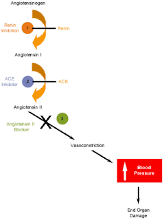
The ONTARGET trial was on the major trials looking at the potential benefits of dual ACE-inhibitor (ACE-I)/angiotensin receptor blocker (ARB) therapy.  Although it was not intended to specifically examine the effects of dual blockade of the renin-angiotensin system on progression of CKD as a primary outcome, a 2008 Lancet article analyzes this trial from a renal perspective.  

ONTARGET was a randomized, double-blind comparison of three different groups:  the ACE-inhibitor ramipril alone (10mg po qd), the ARB telmisartan alone (80mg po qd), or a combination group which received both ramipril and telmisartan.

I won't go into the gory details (a good summary in AJKD by Jeffrey Berns can be found here), but the upshot is that although combination therapy improved proteinuria, it resulted in overall worsened renal outcomes, including a requirement for dialysis or a doubling of serum creatinine.  

There are several caveats to this trial--for example, most of the patients in the trial did not have CKD to begin with, so it is not the ideal population to answer the question of whether or not dual blockade therapy is a good strategy for preventing the progression of existing CKD--but it does suggest that we should not uniformly use the dual-blockade strategy blindly, despite its seemingly sound physiologic rationale.  It also suggests that further trials designed to look at the effects of dual ACE/ARB therapy should look at hard outcomes (such as death, need for dialysis, doubling of serum creatinine, etc) rather than simply using the surrogate marker of proteinuria as a primary outcome. 

Tuesday, February 3, 2009

Kidney Transplant Allocation Policy

I heard a great talk today detailing possible changes in store for the national policy for kidney transplant allocation, as set forward by the Organ Procurement and Transplanation Network (OPTN) (the organization which helps administer UNOS).  

The current allocation system uses a "points" system which is based largely on waiting time:  potential recipients on the list get one point for every year since they are listed.  Points are also given for high senstitivity (4 points for having a PRA >80%) or for having a good match (2 points for a 0 DR antigen mismatch, 1 point for a 1 DR antigen mismatch).  A limited subset of individuals is allowed to "leapfrog" ahead of others in the list:  those with a 0/6 antigen mismatch, those who have donated a kidney previously, or patients <18>

While this system has had some success, there have nonetheless been criticisms aimed at this policy.  One of the major criticisms is that there are frequent instances of mismatch in graft/patient survival--that is, a young individual being given an older kidney (and therefore requiring a second transplant later on down the line) or an old individual being given a younger kidney (and therefore having a high risk of dying with a functional kidney, resulting in several years of "wasted" kidney function).  

Another key criticism of the existing policy is that the SCD/ECD dichotomy is probably too simplistic.  Some ECD kidneys are actually pretty good, and occasionally one will get turned down for use.  Therefore there has been an increased effort to improve the rating system for potential donor kidneys.

The new proposal--which is still being debated and has not actually been formally put forward as a "proposal" per se--significantly deviates from the "point system" in place.  It would be based on the following three ratings:

1.  Donor Profile Index (DPI):  this is the new rating system used to rank the quality of potential donor kidneys.  Instead of classifying the kidney as SCD or ECD, a variety of variables (age, weight, creatinine, etc.) goes into creating a DPI score.  Lower DPI scores indicate a better kidney.

2.  Life Years from Transplant (LYFT):  this measures the difference between expected lifespan WITH a kidney transplant compared to expected lifespan WITHOUT a kidney transplant.  Not surprisingly, young patients tend to have a higher LFYT than, for example, older diabetics.

3.  Dialysis Time (DT):  there is still a component of waiting time which goes into the algorithm--which appeals to a sense of fairness--but the time begins accruing once a patient begins dialysis, as opposed to when one is listed for a transplant.  I think this subtle change is to help eliminate variabilities in health care access (e.g., richer, more educated patients tend to be listed earlier for transplants).  

These three variables combine to create a "Kidney Allocation Score" (KAS) which determines who is matched first.  Overall, the proposed scheme would allegedly lead to better matching of younger donor kidneys with younger recipients (and older donor kidneys with older recipients).  The main criticism of this plan thus far is that it would appear to benefit younger patients and disadvantage older patients.  

Obviously, the proposal does not get around the central problem of transplant nephrology, which is that the organ demand far outweighs the supply.  However, efforts like this to make the organ allocation as efficient as possible (while remaining just and fair) appear to be a good approach to reality.  

More details about the proposed policy changes can be found at the OPTN website.  

Monday, February 2, 2009

Schistosomiasis and Renal Disease

Schistosoma species--also known as blood flukes and bilharzia--can cause some interesting forms of renal disease. Although this is not so common in the United States, it is a very common public health problem in Africa and Asia.

Chronic Schistosoma mansoni infection in about 10-15% cases is associated with a glomerulonephritis. The etiology is not entirely clear but there is evidence that both schistosomal antigens and antibodies against them can be found in glomeruli from biopsy specimens. Similar to the WHO Classification System for lupus, there is a classification system for schistosomal glomerulopathy which has been set forth by the African Association of Nephrology (AFRAN), and there is an interestingly wide variety of pathologies which can result:

-Class I is a mesangial proliferative GN, the recovery from which is generally good.
-Class II is an exudative GN which is classically observed in patients who are co-infected with both Schistostoma as well as Salmonella (apparently common in some regions).
-Class III is an MPGN.
-Class IV is a proliferative sclerosing GN. Classes III & IV are the most common and most severe form of the disease, which may present as an FSGS-like nephrotic syndrome. Concomitant liver disease (common with Schistosomiasis) and IgA deposition are also suggested to play important pathogenic roles.
-Class V is an AA amyloidosis secondary to chronic inflammation from Schistosomiasis.

Schistosoma hematobium, in contrast, is associated with bladder cancer, particularly in the Middle East. Individuals with Schistosomal-induced bladder cancer are generally treated with radical cystectomy with lymph node dissection, with possible roles for adjuvant or neoadjuvant chemotherapy which are still being determined.

Sunday, February 1, 2009

Route of EPO Administration

In the U.S., erythropoietin is typically delivered as 3x/week intravenous injections in the ESRD population. This turns out to be a fairly significant cost, as Medicare-related payments for EPO comprise about 25% of the ESRD budget, which itself is a significant chunk (about 6-7% as of 2002) of the overall Medicare budget.

This cost could probably be decreased by moving to subcutaneous dosing of erythropoietin. One study in France showed in a 1-year crossover study in which ESRD patients received subcutaneous dosing for 6 months, followed by intravenous dosing for 6 months, the EPO dose required to keep the Hgb within a usual target range was far less in the subcutaneous group (74 Units/kg/week) compared to the intravenous group (156 Units/kg/week).

I would imagine that the main barriers towards achieving this would be (a) financial--as it stands now, dialysis units make more money if they administer greater amounts of EPO, (b) legislative--the Social Security Act which has made the Medicare ESRD Program into what it is today specifies that self-administered medications are not covered by Medicare, (c) patient preference--most patients would prefer to avoid another needle stick each dialysis session, and (d) the status quo--that's just the way we're used to doing things.