Monday, January 31, 2011

ARBs and cancer risk?


In the summer of 2010, I came across a health report on the national news that was titled “Angiotensin Receptor Blockers Linked to Cancer”.  The headline was based on a publication that demonstrated an increased cancer signal associated with the use of ARB therapy.  The results appeared in the July 2010 edition of Lancet Oncology.  Riding on the tail of ONTARGET hysteria (see Nate’s article), the results of this study abruptly prompted an FDA safety review on the potential link between ARBs and cancer.  Given the provocative conclusions of this study, I felt it deserved critical review and acknowledgement within RFN.
BACKGROUND
Sipahi et al published “Angiotensin-receptor blockade and risk of cancer: meta-analysis of randomized controlled trials” in July 2010.  The hypothesis for this study was based on experimental studies showing that the renin-angiotensin system, particularly angiotensin II receptors, play an integral role in the regulation of cell proliferation, angiogenesis, and tumor progression.  However, there was a recent study in JCI showing increased longevity in angiotensin II receptor type 1A (AT1A) KO mice and another study in mice showing less tumor growth in AT1A KO mice.
In human studies, the Candesartan in Heart failure Assessment of Reduction in Mortality and Morbidity (CHARM) trial, which assessed ARBs in heart failure, reported an unexpected finding of significantly higher fatal cancers in the candesartan group than with placebo. 
OBJECTIVE
The primary aim of the study was to examine the effect of ARBs on the incidence of new cancer diagnoses.  Secondarily, the study aimed to determine the affect of ARBs on the occurrence of specific solid-organ cancers and mortality related to cancer.  
METHOD
Data was collected using medical search engines.New cancer data were available for 61,590 patients from five trials. Data on common types of solid organ cancers were available for 68,402 patients from five trials, and data on cancer deaths were available for 93, 515 patients from eight trials.
RESULTS
·      Patients randomly assigned to receive ARBs had a significantly increased risk of new cancer occurrence compared with patients in control groups (7.2% vs6.0%, risk ratio [RR] 1.08, 95% CI 1.01–1.15; p=0.016)
·      When analysis was limited to trials where cancer was a pre-specified endpoint, the RR was 1.11 (95% CI 1.04–1.18, p=0.001)
·      Only new lung-cancer occurrence was significantly higher in patients randomly assigned to receive ARBs than in those assigned to receive control (0.9% vs0.7%, RR 1.25, 1.05–1.49; p=0.01)
·      No statistically significant difference in cancer deaths was observed (1.8% vs1.6%, RR 1.07, 0.97–1.18; p=0.183)

LIMITATIONS
·      META-ANALYSIS, not a prospective trial
·      Studies analyzed were not designed or powered to examine cancer incidence or outcomes
·      Cancer data was not available in all trials, suggesting possible publication bias
·      Individual cancer data and timing of cancers was not available
·      The study design precluded adjustment for age, sex, smoking history, etc
·      Most trials used were less than 5 years in duration (too short to adequately assess cancer signal)
In an accompanying editorial, Nissen called for an urgent regulatory review of ARBs and described the study findings as “disturbing”.  Moreover, Nissen stated that ARBs should be used with greater circumspect by the medical community.
Needless to say, the conclusions of this meta-analysis and the accompanying editorial ignited a myriad of assertive retorts criticizing the study design and conclusions.  Experts lashed out against the authors and journal, calling the results “very skewed” and “a bad example of science”.  Interestingly, 10 years ago similar cancer risks were postulated to be associated with amlodipine.  In a subsequent meta-analysis published in the very same journal, Bangalore et al. identified NO excess risk of cancer or cancer death associated with any single anti-hypertensive agent. 
In my opinion, to change practice patterns based on one meta-analysis (that generated data from studies that were never intended to examine cancer occurrences and outcomes) appears immoderate.  The nature of the study design, at best, only suggests a possible link between cancer and ARB therapy.  I have not changed my prescribing pattern based on this study.  What are your thoughts?  

Michael Lattanzio DO

Thursday, January 27, 2011

Orthostatic hypertension and strokes

That's right you read the title correctly- it reads orthostatic hypertension and not hypotension.  Ordering orthostatics on patients is quite common.   Most of the time the clinical query is in regards to the latter (hypotension).  My mentor, Dr. Andreoli, would often refer to performing orthostatics in the hospital as the "death provocation test".  He reasoned that a great majority of hospitalized patients with acute illness will have a significant drop in blood pressure after rising from a seated position.  He felt this was likely not always indicative of volume depletion, but rather from a global sympathetectomy from their acute illness.  If we asked a patient to quickly rise and they did in fact have a robust drop in blood pressure a topple to the floor with resulting head trauma could occur.  Always makes me want to have a couple of extra hands on deck when performing this maneuver.  But, what about performing orthostatic blood pressure measurements on stable outpatients with no complaints. 

I read an interesting editorial in Hypertension this week in regards to orthostatic hypertension.  It took a few glances to read that correctly.  Occasionally I check orthostatic blood pressures on patients in the clinic and get a surprising result.  A huge increase in blood pressure of 40 mmHg from sitting to standing.  I never really thought about the long term consequences of this until I read this editorial and accompanying article by Yatsuya et al.  

Yatsuya et al. prospectively followed 12,817 patients (in the ARIC study) who had baseline orthostatic blood pressure measurements.  They further grouped theses patient into several baseline categories depending on their BP changes from sitting to 2 min after standing.
  1. Decline in BP- Major (-63 to -20), 547 subjects or Minor (-20 to -10), 1507 subjects 
  2. Stable BP- (-10 to +10), 8981 subjects 
  3. Increase in BP- Major (+65 to +20), 303 subjects or Minor (+20 to +10), 1479 subjects
Data was obtained in regards to ischemic stroke incidence and type for a median of 18.7 years.  They report:
  • Little change in BP after standing in the entire cohort.  SBP increased 0.4 mmHg and DBP increased by 3mmHg. 
  • Patients with orthostatic SBP and DBP decreases of greater than 20mmHg had an increased incidence of thrombotic and cardioembolic strokes. 
  • Patients with orthostatic SBP decreases and increases of greater than 20mmHg had an increased incidence of lacunar strokes.  
The authors conclude that these findings are consistent with previous studies showing silent strokes in elderly hypertensive patients with orthostatic hypo- or hypertension.  They speculate that this BP elevation from sitting to standing could be due to excessive sympathetic nervous system activation, which could in turn lead to endothelial dysfunction.  Another theory discussed in the editorial and by these authors is that elderly patients with orthostatic hypertension have been shown have excessive BP dipping at night with greater than a 25mmHg drop in BP (referred to as "extreme dipping").  These "extreme dippers" had an increased risk of silent stroke as well.  Several limitations are present.  Only a baseline orthostatic value is present.  How would these values change over time or would they hold true?  Second, not every participant had an MRI to look for silent strokes.  However, this study was prospective and contains a large sample size.  


How will this change practice? Hard to say.  This could merely serve as a marker for autonomic dysfunction and might not be a modifiable risk factor.  Simple measures such as compression stockings have been used to control both orthostatic hypo- and hypertension.  Beta blockers could be useful in controlling orthostatic hypertension.  Further clinical trials to directly examine these relationships could help in answering these questions.

Wednesday, January 26, 2011

The ECG: QT Interval Abnormalities and Sudden Death in Dialysis Patients

Sometimes overlooked, QT interval abnormalities, namely prolongation and dispersion, are important predictors of sudden death to watch out for in the dialysis unit. Of these two, prolongation of the QT interval is the better studied. The QT interval is a measure of the duration of ventricular de- and re-polarization. Early after-depolarizations, which develop because of a failure of normal repolarization in diseased myocardium or due to certain drugs, result in delayed repolarization and QT interval lengthening. This is an ideal substrate for the development ectopic circuits and arrythymia. Prolonged QT is a well-established risk factor for torsade de pointes, ventricular fibrillation and sudden cardiac death.


Acquired QT prolongation in a dialysis patient has a long list of potential causes, but most important are electrolyte abnormalities (all the hypo’s: hypokalemia, hypomagnesemia, hypocalcemia) and medications. While significant electrolyte abnormalities are (hopefully) rarely missed, as they’re checked and flagged all the time, it is particularly important to pay attention to medications. A study of the prescription claims of almost 5 million patients found the unacceptably high prescription rates of QT-prolonging medications and dual therapy with two or more QT-prolonging drugs. In fact, 0.5% were taking 5 concomitant QT prolonging agents! The paper included 50 medications that prolong the QT interval and 26 that inhibit their hepatic or renal clearance. The commonest culprits are antibiotics (clarithromycin, erythromycin, levofloxacin) and anti-depressants (fluoxetine, amitriptyline, sertraline, venlafaxine).


QT interval dispersion (QTd) is less well recognized and often overlooked. QTd, the longest minus the shortest QT interval on a standard ECG, is a marker of variability in ventricular repolarization. A difference of 50 msec or more has been used as a cut-off in several studies (each small box on the ECG is 40ms). QTd is a risk factor for ventricular arrhythmias and sudden death in the general population. It also appearsto be a useful measure to identify dialysis patients at an increased risk for sudden death, based on the following:


Finally, it is worth remembering that QTd may be an epiphenomenon, as it is associated with LVH and structural heart disease, which are themselves strongly associated with sudden cardiac death in ESRD.

Tuesday, January 25, 2011

Mycobacterial PD peritonitis

One of the most feared complications of peritoneal dialysis is that of PD peritonitis. Infection with gram positive (50-70%) organisms accounts for the vast majority of cases. The typical presentation is with abdominal pain, fever and cloudy dialysate fluid. To make the diagnosis of PD peritonitis, a sample of the PD fluid must be sent for cell count, differential and culture + sensitivity. A PD fluid white cell count of >100/cc is generally accepted as enough for diagnosis.
Empiric therapy can usually be given intra-peritoneally and consists of broad spectrum gram positive and gram negative coverage until the culture data is back. If it is the case where a patient comes in the middle of the night and there is no-one available to give IP antibiotics, then they should be given intravenously.

The cell count differential can sometimes give a clue that pathogens other than bacteria may be lurking around. A predominant lymphocytosis in the PD fluid should raise the suspicion of mycobacterial organisms.

Also, the presence of prolonged symptoms despite treatment of bacterial pathogens, or relapsing peritonitis with negative bacterial cultures, should ring alarms bells and start the search for alternative organisms.

A recent review of tuberculous peritonitis associated with peritoneal dialysis, reported that 89% presented with abdominal pain, 81% with fever and 77% with cloudy dialysate. 30% had a lymphocyte predominant dialysate, but 65% still had a neutrophil predominance. When suspicion arises there are various ways to make the diagnosis – PCR, mycobacterial staining, culture and sometimes a peritoneal biopsy is necessary.

The International Society for Peritoneal Dialysis recommends treatment with isoniazid, rifampin, pyrazinamide and ofloxacin. Three months for pyrazinamide and ofloxacin; with at least 12 months of isoniazid and rifampin. Pyridoxine should be given with isoniazid to reduce the risk of peripheral neuropathy. There is no strong consensus on whether the PD catheter needs to be removed in the case of tuberculous peritonitis – if the patient looks sick, the safer thing to do is remove it, particularly if it is an atypical mycobacterium. In one small study of ten patients, 6 were still on PD nine months after their infection.

TB needs to be kept in the differential for our patients, especially those who are immunosuppressed, come from endemic areas or may have occupational exposures.

Wednesday, January 19, 2011

Better high or low bone turnover disease?

In a recent survey of 630 bone biopsy samples from patients with CKD stage 5 on dialysis, bone turnover was low in 52%, normal in 21% and high in 27% of biopsies. Defective mineralization was found in only 3%. Since the introduction of vitamin D analogs, the dominant bone disease phenotype in dialysis patients has remarkably changed. Now, adynamic bone disease is the primary bone abnormality. Since we are continuously trying to balance vit. D dose/PTH levels, I was interested in the question: if I have to choose, should I error in the side of higher or lower PTH?

I learned some interesting points about bone metabolism that I would like to share with you. When assessing bone volume abnormalities, it is important to differentiate between cortical and cancellous bone. The cortical bone, as the name implies, forms the cortex (outer shell) of most bones, being harder, stiffer and stronger than cancellous bone. It is responsible mainly for the mechanical function of the bones. On the other hand, the cancellous bone typically occurs at the ends of long bones, it has many trabeculations and is highly vascularized. Due to its high surface area, it is the predominant place of metabolic activity of the bone (e.g. exchange of calcium ions).

Now comes the interesting part. Loss of cortical bone occurs mainly in patients with high turnover bone disease, while loss of cancellous bone is often seen in patients with low bone turnover. The clinical outcome of decreased bone strength is fracture, while abnormal metabolic activity results in the inability to maintain mineral homeostasis, which is associated with vascular and soft tissue calcifications. Following these lines, adynamic bone is typically asymptomatic but it is strongly associated with hypercalcemia, cardiovascular calcifications and mortality.

What are the risk factors for adynamic bone disease?
High calcium load, low PTH and vitamin D over-treatment. Older patients, DM and peritoneal dialysis are additional associated factors.

Can we predict the type of bone disease in a dyalisis patient with PTH levels?
No, but it could give you some direction:

  • Intact serum PTH values below 100 pg/mL are associated with a decreased likelihood of osteitis fibrosa and an increased incidence of adynamic bone disease.
  • An intact serum PTH level above 450 pg/mL is typically associated with hyperparathyroid bone disease and/or mixed uremic osteodystrophy.
  • Intermediate PTH levels between 100 and 450 pg/mL may be associated with normal, elevated bone remodeling, or even reduced bone remodeling.

Bone specific alkaline phosphatase levels could also be an adjunctive marker (below 7 ng/mL low bone turnover, above 20 high bone turnover). Radiographic examination of bone can provide important information regarding the presence of hyperparathyroidism (such as subperiosteal resorption). However, radiographic findings are less sensitive than PTH levels and will not establish the type of bone disease. The gold standard is bone bx.

When should you perform a bone bx?
Controversial topic but a bx could be considered in the setting of unexplained fractures, unexplained hypercalcemia, and/or unexplained hypophosphatemia; persistent bone pain; possible aluminum toxicity; and before therapy with bisphosphonates.

How should adynamic bone be treated?
Lower PTH levels should be treated by decreasing the doses of calcium-based phosphate binders, vitamin D, a low dialysate calcium concentration, and perhaps by the use of non-calcium-based phosphate binders (though no data yet supporting this).

My feeling is that we should be cautious in the administration of vitamin D and adjust with small increments based on PTH levels, especially due to the metabolic complications of adynamic bone and difficulty to reverse it. Adynamic bone disease is a relative new entity and long-term complications have not been fully determined yet. I must confess that since PTH does not truly predict the type of bone disease, it is a hell of an empirical area...

Tuesday, January 18, 2011

The Bezold-Jarisch reflex

This intriguing set of clinical signs was discovered by von Bezold and Hirt in 1867 – they found that injection of a veratrum alkaloid caused bradycardia, hypotension and apnea.

In the 1930’s Jarisch and Richter were performing similar experiments in cats to see what effect interruption of the cardiac branches of the vagus nerves would have. They were able to prove that the hypotensive effect described by von Bezold was reflex in origin. Many years later, Dawes was able to prove that the reflex apnoea occurred by a separate mechanism to the haemodynamic changes.

Today the Bezold-Jarisch reflex (BJR) refers to the discoveries of Dawes in 1947 and describes the triad of bradycardia, hypotension and vasodilation that occurs upon stimulation of cardiac receptors.

So, how does all this relate to nephrology? Well, this reflex is an important entity to be aware of when in the haemodialysis unit. To understand why, we need to delve a little bit further into the physiology of the reflex.

Mechanosensitive and chemosensitive receptors in the walls of the ventricles send afferent fibres through the vagus nerve to the vasomotor centres of the brainstem. The basal output from the vasomotor centre is mainly sympathetic, which keeps vessels partially constricted, thereby maintaining blood pressure - this is known as vasomotor tone. The afferent fibres of the BJR have a tonic inhibitory effect on the vasomotor centre, but have a very low rate of basal firing. Upon stimulation, they cause profound inhibition of the vasomotor centre, resulting in decreased sympathetic outflow, bradycardia, hypotension and vasodilation.

Now think of the haemodialysis patient who is 4 or 5 Kg up from their estimated dry weight. As we try to ultrafiltrate the patient, if the rate of fluid removal is greater than their rate of vascular refilling, then there may be trouble ahead. As early hypovolaemia develops, the baroreceptor reflex kicks in and simultaneously the BJR fibres decrease firing – the result is increased sympathetic outflow, increased heart rate and initially, preservation of blood pressure. However, as hypovolaemia becomes more severe, the ventricles contract more vigorously around a poorly filled LV cavity – this is thought to allow the BJR fibres to become paradoxically more active. At this stage the BJR overrides the baroreceptor reflex, causing vasomotor inhibition and the resulting bradycardia, hypotension and vasodilation.

Overall the BJR is felt to be cardioprotective – by causing bradycardia and afterload reduction via peripheral vasodilation, the workload of the heart is reduced and hopefully ischaemia is avoided. See here and here for more information.

I think this is an interesting physiological principle to be aware of in the dialysis unit, where the prevalence of LVH and multiple cardiovascular risk factors is so common. We must closely watch the heart rate and blood pressure during the treatment. Often, tachycardia precedes the precipitous drop in blood pressure – therefore those patients may need closer monitoring and titration of the UF rate.

Thursday, January 13, 2011

Windows to the Soul...and the Vasculature

Continuing the theme of useful Nephrology bedside clinical signs, today we’ll focus on band keratopathy (BK). Often overlooked, this is a band of calcium deposition across the central cornea commonly seen in patients with advanced CKD. It reflects chronic positive calcium balance or hypercalcemia, and is seen in a wide variety of calcium overload states, including myeloma, sarcoid, hyperparathyroidism and renal tubular acidosis. Interestingly, it can regress with treatment of hypercalcemia, (occasionally dramatically, as in this patient) and will often regress following renal transplant too. Calcium is deposited on the corneal surface (directly under the epithelium) as a horizontal band that begins at the periphery and moves centrally as BK becomes more severe. The BK severity grading system reflects this process, with the mildest grades most peripheral (see figure). Deposits begin as a gray haze, progressing to dense white with a pebbly surface. They can lead to pain, a foreign body sensation and recurrent erosions, but are not a cause of visual loss.

The pathophysiology is not fully understood, but passive calcium precipitation is believed to be responsible. Tears and aqueous humor contain calcium and phosphate at concentrations approaching their solubility product. As tears evaporate from the intrapalpebral area, the concentration increases and precipitation occurs. The most severely affected area is the junction of the middle and lower thirds of the cornea, which is the area of maximum atmospheric exposure. Elevated serum calcium increases the likelihood of precipitation occurring. It is worth remembering that a passive precipitation model used to be proposed as the mechanism of vascular calcification in calcium overload states. This has since been debunked; vascular calcification is an active process involving a phenotypic switch in the vascular smooth muscle cell, which comes to resemble an osteoblast. We may yet learn that calcification occurring on the eye is also more complex than just passive deposition.

This last point is relevant as BK and vascular calcification are closely linked: the presence of BK is associated with a ten-fold increased prevalence of vascular calcification, and over half of dialysis patients with BK have radiologic evidence of VC. Furthermore, BK has recently been shown to independently predict 1-year mortality in dialysis patients, with each increment in the BK severity index being associated with a 25% increased mortality risk in multivariate analyses. It would be interesting to study whether BK also predicts calciphyllaxis. When I encounter BK clinically, I tend to treat it as I would extraskeletal calcification at any other site. As it implies chronic positive calcium balance, it prompts me to consider measures to reduce calcium balance in the patient, such as reducing the dialysate calcium bath, switching to a non-calcium containing phosphate binder and/or a non-calcemic vitamin D analog.

Wednesday, January 12, 2011

To ATG or not to ATG?

KDIGO (page S6 of PDF) and the UK renal association have recently published guidelines for the clinical care of kidney transplant recipients. Both of these documents suggest that recipients at ‘higher immunological risk’ should be considered for induction therapy with lymphocyte depleting antibodies (LDAs) such as anti-thymoctye globulin (ATG,)  anti-lymphocyte globulin (ALG) and anti-T cell (CD3) antibody (OKT3).  The US scientific registry of transplant recipients (SRTR) annual report for 2008 states that 44.8% of patients received ATG as induction therapy in that year. As a nephrologist practicing in the UK, I find this surprising. None of the 3 major transplantation centres I have worked at have used LDAs for induction under any circumstances and I have found only a single unit using the drug for induction in the UK; and then only in recipients receiving non-hearting beating kidneys.

This is an intriguing discrepancy and seems to invite the question; which side of the Atlantic is right? I think the question has two parts:
  1. Are LDAs and IL-2 receptor antibodies, the other main class of induction agents (IL2-RAs; such as basiliximab and daclizumab), of comparable efficacy and safety? 
  2. What data justifies preferential use of LDAs in high immunological risk patients as recommended in the guidelines?
A reasonable body of evidence addresses the first question and was summarised in a recent Cochrane review.  With regards to efficacy, patients induced with ATG as compared to IL2-RAs showed less episodes of biopsy-proven rejection at one year but no benefit to rates of graft loss or clinically diagnosed or steroid-resistant rejection. Concerning safety, the review found significant reductions in CMV disease and malignancy when IL2-RAs were used over LDAs. The authors derived a number needed to treat of 16 to avoid one case of CMV disease and of 58 to prevent one case of cancer.

The evidence base for question 2 has been accumulating since at least 1998 when a meta-analysis showed that LDA induction mediated reduction in graft loss (compared to placebo) was greater in patients with high panel-reactive antibody. Since then, two recent high-quality randomised controlled trials (Brennan et al. and Noel et al.) have been performed specifically in high risk recipients. The trials addressed diverse patient populations and used different IL2-RAs and maintenance regimes but produced similar findings of significant reductions in rejection rates at one year without differences in graft or patient survival (although Brennan et al. used a composite end point).

So, the recommendations about the use of ATG in high-risk patients are based on decreased rates of rejection. However, decreased rejection has not translated into increased graft survival in a number of studies looking at ATG vs IL2-RAs (summarised in this editorial).

Having looked through the evidence I feel that almost 45% ATG induction is too high, and the UK’s avoidance of ATG probably unwise too. I think the next challenge would be to produce an evidence based decision algorithm for assigning patients into high and low risk groups. Anybody else have any thoughts?

Tom Oates, MD

Monday, January 10, 2011

Role of erythropoietin in acute kidney injury: what does the evidence say?

Since the discovery of Erythropoietin in 1977 and subsequent cloning of the gene in 1985, recombinant Erythropoietin (EPO) has been widely used in the management of anemia due to cancer, chemotherapy, and chronic kidney disease.

Although the endocrine effects of EPO in stimulating maturation and differentiation of erythroid precursor cells in the bone in response to hypoxia is widely known, the anti-apoptotic and anti-inflammatory properties particularly in the critical organs such as kidney, heart and brain were discovered only in the last decade.

Researchers from the United States demonstrated that EPO receptors are expressed in the renal tubular epithelial cells, proximal tubular cells, and mesangial cells. EPO induced activation of these receptors leads to activation of Janus activated kinase 2(JAK-2) pathway which in turn stimulates several other signaling pathways (MAPK,NFKB,STAT 3/5), all of which promote anti-apoptotic and proliferative proteins that increase cell survival (read this article).

With this knowledge, researchers from Australia demonstrated that EPO administration both in vitro and in vivo in doses up to 5000 U/kg in animals with ischemic AKI hastened cell recovery and prevented cell death. Several other researchers have demonstrated this effect in different injury models (hypoxia, ischemia-reperfusion, nephrotoxins, sepsis etc).
As would be expected, these promising results in animal studies led to a randomized, double blind, placebo controlled trial (EARLYARF) in 529 ICU patients who were identified at risk of AKI by urinary biomarkers(c-glutamyl transpeptidase and alkaline phosphatase ≥46.3). Administration of 100,000 units of EPO IV in 2 divided doses over 24 hrs did not reduce the risk of developing AKI, quite contrary to what was seen in animal studies.
In my opinion, likely causes for this discrepancy included:
1) Dosing in animals had been much higher (equivalent to up to 350000 units in humans).
2) Optimal timing of EPO administration might be an issue (within 6 hrs of injury)
3) Unclear, if the ideal urinary biomarkers were used.

Further studies with proper dosing and timing of EPO administration and early identification of patients with risk of AKI using ideal urinary biomarkers may answer this question best. Besides, the utility of administering EPO to ICU patients to reduce transfusions has already been refuted, is not cost effective and may be associated with 40% increased risk of thrombotic events (read this blog). This leaves us with no good indication to consider EPO in critically ill patients with AKI (not ESRD).

If you are a big believer of basic research, you may still want to consider EPO for its favorable actions, but with recent negative publicity EPO has been receiving, it appears like the clock is quickly moving towards “may be not or even no”.

Saturday, January 8, 2011

Jugular venous distension - clinical studies

In the previous blog we discussed some of the physiological principles underlying the JVP. Now I thought I’d present some manuscripts relating to the utility of the isolated use of JVP in clinical practice.

The first is a comparative study of hand-held ultrasound examination of IVC diameter vs clinical JVP assessment in determining RA pressure among medical residents. All assessments occurred within one hour of patients having a right heart cath. One resident, blinded to the ultrasound results performed the clinical examination in 40 patients. The JVP was not visualized in 37% of the 40 patients. In the remaining 63%, the sensitivity for predicting an RA pressure >10 mm Hg was 82% with ultrasound and 14% from JVP inspection. This of course is an extremely small study, with only one person at one experience level performing the exam.

An interesting study examining the presumed distance between the sternal angle and the mid right atrium recruited 160 patients undergoing CT chest scans. Using geometric calculations, they estimated the sternal angle - RA distance to be 8 cm, 9.7 cm and 9.8 cm at 30, 45 and 60 degrees elevation respectively. They found considerable inter-subject variability, with dependent variables including age, smoking status and AP chest diameter. This really emphasizes the need for a standard reference point to allow intra-patient comparisons and poses questions of the ability to compare similar measurements in different patients.

A systematic review published in Chest in 2008 examined the usefulness of measured CVP in relation to blood volume, CVP in determining fluid responsiveness and finally delta CVP in determining fluid responsiveness. 24 studies with a total of 803 patients were included. The pooled correlation coefficient between CVP and blood volume was 0.16 (95% CI 0.03, 0.28). Following fluid administration, the pooled correlation coefficient between baseline CVP and delta Cardiac Index was 0.18 (95% CI 0.08, 0.28); the pooled correlation coefficient between delta CVP and delta Cardiac Index was 0.11 (95% CI 0.015, 0.21). These results suggest a poor relationship between the isolated inspection of CVP and prediction of blood volume and fluid responsiveness, with the caveats of study heterogeneity in a systematic review.

These studies underscore that the JVP should not be used in isolation, as a measure of volume status nor fluid responsiveness. Furthermore, in order to derive any usefulness from it, we must understand what it actually signifies. As long as we perform it with a standard approach in each patient, understanding the fact that it’s a window to the pressure in the RA (not volume) and if it is part of an overall patient examination, then I think it can help with a detailed clinical work-up. Remembering that there are many processes that can cause an elevated JVP will help broaden your differential and keep you alert to the possibility of alternative patho-physiological processes. As one of my attendings said, 'it's free and you can do it everyday'.

Wednesday, January 5, 2011

TRAPS

It’s been a long time since my last blog post, so I have decided to catch up by writing about two cases I saw in my transplant clinic. However, they have the same disease, a brother and sister who inherited the same autosomal dominant disease called tumor necrosis factor (TNF) receptor-1 associated periodic syndrome (TRAPS, formerly known as familial Hibernian fever). The genetic defect in TRAPS resides in the gene that encodes the 55 kDa receptor for tumor necrosis factor, TNFR1. These mutations lead to loss of normal function rather than gain of function as you may think, and thus the pathogenesis of TRAPS is an enigma. A recent PNAS publication shed some light on the pathogenesis of the disease. They showed that mutant TNFR1 accumulates intracellularly in peripheral blood mononuclear cells of TRAPS patients and in multiple cell types from two independent lines of knock-in mice harboring TRAPS-associated TNFR1 mutations. Mutant TNFR1 did not function as a surface receptor for TNF but rather enhanced activation of MAPKs and secretion of proinflammatory cytokines. These patients will present with recurrent fevers over months or years, in the absence of associated viral or bacterial infections. They also may have focal or sometimes migratory myalgias, conjunctivitis, periorbital edema, abdominal pain, monoarticular arthritis, and rash.
You might be wondering how they ended up in my transplant clinic. The answer is secondary (AA) amyloidosis primarily involving the kidney. However it only occurs in a minority of patients (approximately 15 percent in the United States).
Managing patients with TRAPS consists mainly of Etanercept, a fusion protein consisting of two copies of the 75kDa TNF receptor (TNFR2) bound to the Fc portion of human IgG. Etanercept can be highly effective in many patients with TRAPS, though not all respond and the response is sometimes partial.
Finally, the major question remains, do those patients need to be on Etanercept after transplantation, considering that they are already immunosuppressed with our maintenance regimen? Well, it’s not clear in the literature…. In our cases, the brother was maintained on Etanercept and he didn’t have any flare of the disease with stable kidney function and no recurrence of amyloidosis. The sister however, was not on Etanercept and she had recurrence of AA amyloidosis in the transplanted kidney. My approach will be to keep those patients on Etanercept and have low threshold to reduce maintenance immunosuppression.

Tuesday, January 4, 2011

Jugular venous distention- the physical exam, continued...

Following on from Finnian's last post, here are some remarkable videos from the 1950's on JVP examination (physical examination of JVP begins at 3:38):

Monday, January 3, 2011

Intradialytic hypertension

I’ve recently had a few calls from our outpatient dialysis unit about patient’s with rises in their blood pressure during and after dialysis. Intradialytic hypotension is a very common phenomenon and it’s been interesting to learn that intradialytic hypertension also occurs quite frequently with a reported prevalence of up to 15%.

The relationship between blood pressure and clinical outcome in hemodialysis patients is complex and both the most predictive type of measurement and optimal target value are unknown. As a matter of practicality, The Handbook of Dialysis recommends targeting pre dialysis blood pressures of <130/80 with less strict goals in patients with wide pulse pressures or at risk for orthostasis. Intradialytic hypertension has been defined in a number of ways but basically refers to patient’s whose intradialysis or post dialysis BPs are higher than their starting values. It has been associated with increased hospitalization and mortality in hemodialysis patients.

Why it occurs is unknown but several theories have been put forward:

1) Volume overloadAnimal chronic kidney disease models suggest that chronic sodium and volume overload can lead to sustained elevations in blood pressure though increases in peripheral vascular resistance. On the molecular level sodium leads to the release of digitalis-like factor which inhibits the Na/K ATPase on vascular smooth muscle leading to increases in both intracellular sodium as well as calcium. These rises in intracellular calcium then cause smooth muscle contraction with increases in vascular resistance. In addition, dialysis patients with little or no residual renal function are not able to urinate off excess volume leading to rises in preload and cardiac output.

So the above provides an explanation as to why sodium mediated volume overload might lead to hypertension but why would blood pressure rise further with ultrafiltration on dialysis?

Some studies suggest that as volume is removed in overloaded patients cardiac output and blood pressure rise, presumably as the heart is put in a more favorable portion of the starling curve though evidence for this is conflicting. Alternatively a recent review tied together the volume overload and endothelin hypotheses (see below) by suggesting that faster intravascular refilling in volume overloaded patients resulted in more mechanical stress triggered endothelin-1 release with subsequent rises in peripheral vascular resistance.

2) Endothelial cell dysfunction – In response to volume changes, mechanical stress and hormonal stimuli endothelial cells synthesize and release factors that contribute to BP homeostasis. Severalstudies to date have looked at this and have shown greater rises in the vasoconstrictor endothelin-1 in patients with intradialytic hypertension when compared with controls. The largest study supporting this hypothesis also showed that intradialytic rises in blood pressure were largely due to rises in peripheral vascular resistance rather than cardiac output. Of interest, there is a currently enrolling NIH trial looking at carvedilol's impact on intradialytic hypertension due to it's potential to suppress endothelin-1 release.

3) Dialytic removal of antihypertensives – Many blood pressure medications including multiple beta-blockers and ACE inhibitors are significantly removed during dialysis. Although removal of these agents is a potential contributor in some cases, intradialytic hypertension still occurs in patients who are off all BP meds.

4) Erythropoetin stimulating agents – Intravenous administration has been associated with elevations in blood pressure in dialysis patients and interestingly also with elevations in endothelin-1.

5) Sympathetic overactivity, RAAS activation, electrolytes – Volume removal followed by upregulation of homeostatic systems is often cited as a possible cause of intradialytic hypertension. However, a recent study that measured plasma catecholamines and renin in patients with and without intradialytic hypertension pre and post dialysis found that renin and norepinephrine were actually higher in controls post dialysis. Dialysis induced reductions in serum potassium and elevations in calcium have also been postulated as possible causes but in the same study no significant difference between groups in these electrolytes was found. Intradialytic sodium gain due to higher dialysate than plasma sodium has been suggested as a possible cause of intradialytic hypertension but has not been directly studied.

Interventions targeting improvements in intradialytic hypertension have not been evaluated in randomized prospective fashion and the optimal treatment approach and benefit, if any, is unknown.

When I get the phone call I’ve been making sure the patient is asymptomatic then depending on the details of the blood pressure, I ask the patient and nursing staff if I can extend the run and increase the ultrafiltration goal (two small case series support this approach). If the BP remains elevated after dialysis, I will ask the patient to take an additional dose of one of their antihypertensives and I make note to review their medications, epo dosing, labs, dialysis regimen and dry weight.

I’d be interested to hear how others deal with this dilemma.

Sunday, January 2, 2011

Jugular venous distention- the physical exam

The physical examination of volume status is perhaps the most common assessment we perform in everyday clinical practice. It is also one of the most difficult and subjective tasks to perform. In particular, the JVP is one of these mystical signs that even very accomplished physicians can find hard to get right. So, here are some essential facts about the JVP that the trainee nephrologist needs to know:

- The JVP should ideally be measured on the right side using the internal jugular vein, as anatomically this is the straightest column of blood in contact with the right atrium
- The JVP gives an indication of the pressure in the right atrium – it is not a direct measure of volume. We can infer some information regarding the volume status based on the pressure, but must remember that there are other influences at play.
- The JVP should be measured with the patient at 45 degrees to the horizontal. In this position, the sternal angle is a vertical distance of ~5cm above the right atrium
- By convention the JVP is measured as the vertical height from the sternal angle, but many people add the additional 5cm when reporting it – the important thing is to state the reference point, i.e. RA or sternal angle
- A measurement of >3cm from sternal angle (>8cm from RA) is taken as evidence of high RA pressure in normal patients
- Points helpful in distinguishing the JVP from the carotid:
Visible, not palpable
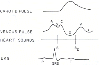
Complex waveform – see here for more details
Varies with respiration – usually decreases on inspiration
Fills from above
Increases with pressure on the abdomen – the hepatojugular reflux

Most importantly, there are many conditions that can result in an elevated JVP:
1. Right ventricular failure
2. Tricuspid regurgitation or stenosis
3. Pericardial effusion or constrictive pericarditis
4. SVC obstruction – usually no waveform as transmission from the RA is blocked
5. Volume overload
In the next post I’ll try to cover some of the previous studies that have the examined the use of the JVP in clinical assessments and trials. Hopefully this has provided some useful review for trainees in one of the common everyday clinical practices.