Showing posts with label physical exam. Show all posts

Wednesday, January 4, 2012

Is the glass half empty or half full?

Recently, we were asked to see a patient on the consult service with a rising creatinine, a BUN of close to 200 and decreased urine output. The patient was a diabetic who had an STEMI several months previously complicated by pneumonia and cardiogenic shock. He was cachectic, did not have any edema or ascites and because of his trach-collar, I put “unable to assess JVD” in my note. His BUN had been high (above 100) for more than 4 weeks (not on TPN, steroids, no GI bleeding) as he was aggressively diuresed for heart failure and his creatinine had been rising slowly which, given his low muscle mass, indicated a significant reduction in GFR. My attending also assessed the patient and felt that his JVP was elevated and recommended diuresis. The primary team, in contrast, felt that his JVP was low and they recommended fluids. The assessment of the JVD is a notoriously difficult exam and very hard to master if one does not do it appropriately as described in a previous post. The patient received 2 liters of fluid and his cardiac status worsened so he was started on dialysis several days later for worsening renal function and volume removal.

This case illustrates the difficulty in accurately assessing volume status in patients in general.

When was the last time you assessed a JVD comfortably? We often say "this patient is dry" or that he needs diuresis but how accurate are our assessments based on the clinical exam alone?

A previous post discussed the JVP and its use as a tool for volume status assessment and cited a systematic review stating that there is a “poor relationship between the isolated inspection of CVP and prediction of blood volume and fluid responsiveness.” One of my attendings gave me an article on this topic several weeks ago: "Clinical assessment of extracellular fluid volume in hyponatremia". The article assessed the clinical judgment of volume status by one of the authors (including cardiac parameters, JVP, orthostatic changes, skin turgor, moisture in the axillae, hydration of mucous membranes) and volume status was 'objectively' assessed by spot urine samples of sodium and creatinine and BUN, norepinephrine and plasma renin concentrations. The clinical assessment was only able to identify 47 % of hypovolemic patients and 48% of normovolemic patients whereas the spot urine sodium clearly separated hypovolemic from normovolemic patients.

The "Bible" of physical examination - Evidence Based Physical Diagnosis by Steven McGee - attributes a low sensitivity or specificity or both to the most common findings used when assessing hypovolemia (the highest likelihood ratio was 2.8 for a dry axilla; in contrast, dry mucous membranes, tongue furrows, sunken eyes, confusion, weakness or unclear speech did not have a significant likelihood ratio). Capillary refill time has been compared only once to a diagnostic standard and was found to have no diagnostic significance.

An intriguing series in JAMA about the rational physical exam stated in the conclusion that "in patients with vomiting, diarrhea, or decreased oral intake, few findings have proven utility, and clinicians should measure serum electrolytes, serum blood urea nitrogen, and creatinine levels when diagnostic certainty is required."

The bottom-line I learned from all of this is: our examination at the bedside is notoriously unreliable in making accurate statements about a patient's volume status and objective parameters need to be taken into account to get a complete picture. A previous post discussed the use of urine electrolytes as a more objective tool for assessing volume status in addition to clinical examination.

Posted by Florian Toegel

Tuesday, January 18, 2011

The Bezold-Jarisch reflex

This intriguing set of clinical signs was discovered by von Bezold and Hirt in 1867 – they found that injection of a veratrum alkaloid caused bradycardia, hypotension and apnea.

In the 1930’s Jarisch and Richter were performing similar experiments in cats to see what effect interruption of the cardiac branches of the vagus nerves would have. They were able to prove that the hypotensive effect described by von Bezold was reflex in origin. Many years later, Dawes was able to prove that the reflex apnoea occurred by a separate mechanism to the haemodynamic changes.

Today the Bezold-Jarisch reflex (BJR) refers to the discoveries of Dawes in 1947 and describes the triad of bradycardia, hypotension and vasodilation that occurs upon stimulation of cardiac receptors.

So, how does all this relate to nephrology? Well, this reflex is an important entity to be aware of when in the haemodialysis unit. To understand why, we need to delve a little bit further into the physiology of the reflex.

Mechanosensitive and chemosensitive receptors in the walls of the ventricles send afferent fibres through the vagus nerve to the vasomotor centres of the brainstem. The basal output from the vasomotor centre is mainly sympathetic, which keeps vessels partially constricted, thereby maintaining blood pressure - this is known as vasomotor tone. The afferent fibres of the BJR have a tonic inhibitory effect on the vasomotor centre, but have a very low rate of basal firing. Upon stimulation, they cause profound inhibition of the vasomotor centre, resulting in decreased sympathetic outflow, bradycardia, hypotension and vasodilation.

Now think of the haemodialysis patient who is 4 or 5 Kg up from their estimated dry weight. As we try to ultrafiltrate the patient, if the rate of fluid removal is greater than their rate of vascular refilling, then there may be trouble ahead. As early hypovolaemia develops, the baroreceptor reflex kicks in and simultaneously the BJR fibres decrease firing – the result is increased sympathetic outflow, increased heart rate and initially, preservation of blood pressure. However, as hypovolaemia becomes more severe, the ventricles contract more vigorously around a poorly filled LV cavity – this is thought to allow the BJR fibres to become paradoxically more active. At this stage the BJR overrides the baroreceptor reflex, causing vasomotor inhibition and the resulting bradycardia, hypotension and vasodilation.

Overall the BJR is felt to be cardioprotective – by causing bradycardia and afterload reduction via peripheral vasodilation, the workload of the heart is reduced and hopefully ischaemia is avoided. See here and here for more information.

I think this is an interesting physiological principle to be aware of in the dialysis unit, where the prevalence of LVH and multiple cardiovascular risk factors is so common. We must closely watch the heart rate and blood pressure during the treatment. Often, tachycardia precedes the precipitous drop in blood pressure – therefore those patients may need closer monitoring and titration of the UF rate.

Saturday, January 8, 2011

Jugular venous distension - clinical studies

In the previous blog we discussed some of the physiological principles underlying the JVP. Now I thought I’d present some manuscripts relating to the utility of the isolated use of JVP in clinical practice.

The first is a comparative study of hand-held ultrasound examination of IVC diameter vs clinical JVP assessment in determining RA pressure among medical residents. All assessments occurred within one hour of patients having a right heart cath. One resident, blinded to the ultrasound results performed the clinical examination in 40 patients. The JVP was not visualized in 37% of the 40 patients. In the remaining 63%, the sensitivity for predicting an RA pressure >10 mm Hg was 82% with ultrasound and 14% from JVP inspection. This of course is an extremely small study, with only one person at one experience level performing the exam.

An interesting study examining the presumed distance between the sternal angle and the mid right atrium recruited 160 patients undergoing CT chest scans. Using geometric calculations, they estimated the sternal angle - RA distance to be 8 cm, 9.7 cm and 9.8 cm at 30, 45 and 60 degrees elevation respectively. They found considerable inter-subject variability, with dependent variables including age, smoking status and AP chest diameter. This really emphasizes the need for a standard reference point to allow intra-patient comparisons and poses questions of the ability to compare similar measurements in different patients.

A systematic review published in Chest in 2008 examined the usefulness of measured CVP in relation to blood volume, CVP in determining fluid responsiveness and finally delta CVP in determining fluid responsiveness. 24 studies with a total of 803 patients were included. The pooled correlation coefficient between CVP and blood volume was 0.16 (95% CI 0.03, 0.28). Following fluid administration, the pooled correlation coefficient between baseline CVP and delta Cardiac Index was 0.18 (95% CI 0.08, 0.28); the pooled correlation coefficient between delta CVP and delta Cardiac Index was 0.11 (95% CI 0.015, 0.21). These results suggest a poor relationship between the isolated inspection of CVP and prediction of blood volume and fluid responsiveness, with the caveats of study heterogeneity in a systematic review.

These studies underscore that the JVP should not be used in isolation, as a measure of volume status nor fluid responsiveness. Furthermore, in order to derive any usefulness from it, we must understand what it actually signifies. As long as we perform it with a standard approach in each patient, understanding the fact that it’s a window to the pressure in the RA (not volume) and if it is part of an overall patient examination, then I think it can help with a detailed clinical work-up. Remembering that there are many processes that can cause an elevated JVP will help broaden your differential and keep you alert to the possibility of alternative patho-physiological processes. As one of my attendings said, 'it's free and you can do it everyday'.

Tuesday, January 4, 2011

Jugular venous distention- the physical exam, continued...

Following on from Finnian's last post, here are some remarkable videos from the 1950's on JVP examination (physical examination of JVP begins at 3:38):

Sunday, January 2, 2011

Jugular venous distention- the physical exam

The physical examination of volume status is perhaps the most common assessment we perform in everyday clinical practice. It is also one of the most difficult and subjective tasks to perform. In particular, the JVP is one of these mystical signs that even very accomplished physicians can find hard to get right. So, here are some essential facts about the JVP that the trainee nephrologist needs to know:

- The JVP should ideally be measured on the right side using the internal jugular vein, as anatomically this is the straightest column of blood in contact with the right atrium
- The JVP gives an indication of the pressure in the right atrium – it is not a direct measure of volume. We can infer some information regarding the volume status based on the pressure, but must remember that there are other influences at play.
- The JVP should be measured with the patient at 45 degrees to the horizontal. In this position, the sternal angle is a vertical distance of ~5cm above the right atrium
- By convention the JVP is measured as the vertical height from the sternal angle, but many people add the additional 5cm when reporting it – the important thing is to state the reference point, i.e. RA or sternal angle
- A measurement of >3cm from sternal angle (>8cm from RA) is taken as evidence of high RA pressure in normal patients
- Points helpful in distinguishing the JVP from the carotid:
Visible, not palpable
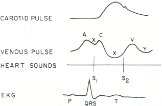
Complex waveform – see here for more details
Varies with respiration – usually decreases on inspiration
Fills from above
Increases with pressure on the abdomen – the hepatojugular reflux

Most importantly, there are many conditions that can result in an elevated JVP:
1. Right ventricular failure
2. Tricuspid regurgitation or stenosis
3. Pericardial effusion or constrictive pericarditis
4. SVC obstruction – usually no waveform as transmission from the RA is blocked
5. Volume overload
In the next post I’ll try to cover some of the previous studies that have the examined the use of the JVP in clinical assessments and trials. Hopefully this has provided some useful review for trainees in one of the common everyday clinical practices.