Friday, December 29, 2017

The ELITE & the Rest in Kidney Transplantation

Image result for cocktail party
The ELITE-Symphony study enshrined standard triple therapy immunosuppression of tacrolimus, mycophenolic acid and prednisolone, for renal transplant recipients. We get familiar with these agents relatively quickly in our renal training, but what about the other less common agents that are used?  Why do we not use them as first line, when should we consider them and what side effects do we need to be aware of?
Sirolimus and everolimus are mTOR inhibitors (see previous RFN post), and prevent cell cycle progression and lymphocyte proliferation.  Their anti-proliferative effects mean they have a role in conditions such as tuberous sclerosis, psoriasis and certain cancers.  The antiproliferative and antiangiogenic effects pose issues post-surgery however with poor wound healing and lymphocele formation meaning they should not be used for around 6 months post-transplant.  Other side effects include pneumonitis, hyperlipidaemia, bone marrow suppression, thrombotic microangiopathy and proteinuria due to FSGS-type lesions. Alongside this relatively long list of side effects, the main concern with sirolimus is related to mortality. A meta-analysis of 21 studies with a total of 5876 patients showed sirolimus was  associated with increased mortality post-transplant, mainly from cardiovascular and infectious complications (adjusted hazard ratio 1.43, 95% CI 1.21-1.71). So why do some patients end up on sirolimus?  The same meta-analysis showed that sirolimus is associated with a 40% reduced risk of malignancy, particularly in non-melanoma skin cancers. The most common reasons for switching to mTORi are malignancy, often recurrent skin cancers, and for calcineurin-inhibitor (CNI) induced injury (although if eGFR<40mls/min or overt proteinuria outcomes are likely to be poor or no better with a switch). Moreover, as seen in ELITE-Symphony and other, acute rejection rates and patient dropouts are consistently higher with mTORi compared to CNIs.
Image result for belatacept mechanismAnother agent we may be hearing more about in the future is belatacept: a co-stimulation pathway blocker. It is attractive as it is designed to replace the CNI in the treatment regime. It binds to CD80 and CD86 on antigen-presenting cells, thereby blocking CD28 mediated activation of T Cells. It is administered as an IV infusion every 4-6 weeks alongside steroid and an anti-metabolite.
The BENEFIT and BENEFIT-EXT (extended criteria donors) trials compared high and low intensity belatacept regimes with cyclosporine in non-sensitised recipients undergoing DBD or living donor renal transplants. Over 7 years of follow-up, they found improved patient and graft survival with belatacept in BENEFIT and better measured GFR and reduced formation of DSA with belatacept in both studies. There was an increased incidence of acute rejection in BENEFIT although this obviously didn’t translate into inferior outcomes. There were also initial concerns regarding a small increase in cases of PTLD in EBV seronegative recipients although longer term data was reassuring that the overall risk of this was very low.
A potential benefit of belatacept is it’s a directly observed therapy, so compliance will be known. Although it is expensive, if patients experience improved graft outcomes then any longer term savings need to be considered. However what about patients who are highly sensitised? Many patients we want to avoid CNIs in are sensitised but here is a dearth of data in using belatacept in this circumstance. Moreover, the studies compared belatacept to cyclosporine, not tacrolimus, the current standard of care. Currently belatacept is not available for new patients due to supply issues, another issue likely preventing its more widespread use.  
Post by Ailish Nimmo

Sunday, December 24, 2017

Adenovirus and graft versus host disease in the kidney

A young man with a myelodysplastic syndrome status post allogeneic bone marrow transplant was recently found to have graft-versus-host disease (GVHD) of the skin and presumed liver involvement which was treated with tacrolimus and methotrexate. The increased immunosuppression was complicated by an upper respiratory tract infection which was treated with empiric cefepime; also, at that time,  a nasopharyngeal swab culture tested positive for rhinovirus.

The patient was discharged on amoxicillin-clavulanate to complete a 14-day course. A few days later, the patient presented to the hospital with dysuria and hematuria. Kidney function was within normal limits (creatinine 0.6-0.8 mg/dL) on admission and he was diagnosed with rhinovirus infection again. Additionally,  he was found with a concomitant adenovirus infection, confirmed with culture and DNA PCR amplification test (2,250,000 copies/ml). Also, the patient developed acute kidney injury (AKI) with a rise in creatinine up to 1.32 mg/dL. He had 5.6 grams of proteinuria on 24 hour urine collection and he was presumed to have adenovirus related hemorrhagic cystitis.

A renal biopsy was performed for further management of the acute kidney injury, revealing signs of both GVHD and adenovirus infection. In order to treat the patient’s GVHD of the native kidney he was given methylprednisolone and was continued on tacrolimus. Furthermore, for the hemorrhagic cystitis due to adenovirus infection, he was given intravesicular and intravenous cidofovir, probenecid, in addition to hyperbaric oxygen. The patient’s kidney function improved, however, he had recurrent AKI due to obstructive nephropathy due blood clots, for which he underwent bilateral stent placement. At the time of discharge, the patient was hemodynamically stable and kidney function was almost at his baseline.

According to the National Institute of Health (NIH) consensus criteria, Acute GVHD usually happens within the first 100 days after transplant, then it becomes chronic.  Classification is based mostly on clinical findings, rather than period of time. Acute GVHD is characterized by maculopapular rash, nausea and vomiting, diarrhea, ileus or hepatitis;  persistent, recurrent, or late-onset GVHD have symptoms of acute GVHD but it presents after 100 days, without features of chronic GVHD. Overlap GVHD happens when the patient has both symptoms of acute and chronic GVHD. It has been reported that patients with overlap syndrome have increased morbidity and mortality. GVHD rarely affects the kidney and usually presents after decreasing immunosuppressive therapy. In the chronic form it usually causes nephrotic syndrome such as membranous glomerulonephropathy and minimal change disease; less commonly it can cause focal segmental glomerulosclerosis, IgA nephropathy, and membranoproliferative glomerulonephritis.

First line therapy includes corticosteroids either topical or intravenous, depending on the stage. When the GVHD is grade I limited to the skin, it is treated with topical corticosteroids and possible adjustments of the patient's methotrexate and tacrolimus . Grade II GVHD or higher is treated with high-dose systemic corticosteroids- usually 2 mg/kg/day of intravenous methylprednisolone or oral steroids. Finally, extensive GVHD is treated with high-dose systemic corticosteroids, and occasionally with cyclosporine with trough levels of 200-450 ng/ml. Tacrolimus has been used in some case reports.

Adenovirus may cause an upper respiratory tract infection. It can cause a tubulointerstitial nephritis and hemorrhagic cystitis which affects the kidney as well, usually seen after a renal transplant. Diagnosis can be made by viral culture, viral antigen assay and PCR. Renal biopsy is needed for definitive diagnosis of renal involvement. On light microscopy, we can see necrotizing granulomas with interstitial nephritis and electron microscope shows icosahedral virions that form large paracrystalline aggregates with the nuclei of infected cells. Current treatment is with cidofovir with probenecid to decrease its nephrotoxic effects. Concurrently, if the patient is on immunosuppressive therapy, it should be decreased to promote immune recovery. Hyperbaric oxygen has been shown to treat radiation and cyclophosphamide induced hemorrhagic cystitis, and according to several small case reports, it has been used to treat adenovirus-induced hemorrhagic cystitis.

In conclusion, this is a unique case not commonly encountered in which a patient suffered from adenovirus infection and GVHD, concomitantly. Renal biopsy is generally required for diagnosis and when both diseases are present, there is a challenge on how to manage immunosuppression.

Cassiopia Lippold, MD
Second year Nephrology Fellow
University of Maryland Medical Center

Friday, December 15, 2017

Another Great Masquerader: Amyloidosis

A middle-aged man with a history of microcytic anemia, well-controlled diabetes mellitus (DM)/hypertension (for 15 years), and a 16 month history of diarrhea with a 30-pound weight loss, and a creatinine of 1.9 mg/dL.

Laboratory tests showed a urine protein to creatinine ratio of 0.35 mg/g, hemoglobin of 9.3 g/dL, white blood cell count of 13.5 x 10^9/L, and a platelet count of 662 x 10^9/L. A workup for chronic diarrhea revealed an elevated erythrocyte sedimentation rate and C-reactive protein of 85 mm/hr and 18 mg/dL, respectively. Serologies for systemic lupus erythematosus, mixed connected tissue diseases, tuberculosis, hepatitides, and monocolonal gammoapthies were also unrevealing. Computed tomography of the abdomen, EGD, and colonoscopy were unrevealing – biopsy specimens of both the small and large bowel did not show evidence of inflammation. His urine sediment did not have red blood cells (RBCs) or RBC casts.

Despite his long-standing DM and hypertension, a kidney biopsy was performed – which revealed AA amyloidosis (see immunohistochemistry staining for amyloid A protein and an electron microscopy images below)


Endoscopic biopsies were subsequently stained for amyloid A protein – which was also positive.

Serum amyloid A (SAA) is part of a family of apolipoproteins associated and an acute phase reactant whose production is upregulated by the liver typically in the setting of infection or inflammation, which leads to increased expression of inflammatory cytokines like interleukin (IL)-1, IL6, and TNF-alpha. Increased SAA levels ultimately lead to the formation of amyloid fibrils that are immune to proteolysis.

The differential diagnosis of AA amyloidosis is broad – including autoimmune disorders, inflammatory arthritis, inflammatory bowel disease, lymphoma, familial Mediterranean fever, IgG4-related disease, Castleman disease, and chronic infection. A study of the natural history and outcome in systemic AA amyloidosis of a few hundred patients found that the underlying disorder was inflammatory arthritis in 60%, chronic sepsis in 15%, and periodic fever syndromes in 9%. The underlying disorder was unknown in 6% of cases.

Like most other secondary diseases processes, treatment for AA amyloidosis is treatment of the underlying disease. Other therapies should enhance clearance of amyloid deposits, decrease or interrupt fibril assembly, and decrease fibril deposition. For example, two agents that have been used with some success are a monoclonal antibody to IL-6 (tocilizumab) and the newer agent eprodisate, which directly interferes with amyloid fibril formation and deposition. SAA levels may predict disease progression and prognosis. These therapies would be potential options in this patient who did not have a proven underlying disease process.

To conclude, the presentation of amyloidosis can be nonspecific and the differential is widespread. It is important to remember that kidney biopsy can change management in a significant percentage of cases – even in a case like this, where the urinalysis was misleading and masked underlying amyloidosis.

*This is a fictionalized case based on a true account, details have been modified/changed

Samira Farouk, MD
Chief Fellow, Division of Nephrology
Icahn School of Medicine at Mt. Sinai


Vote for the Top Nephrology-Related Stories of 2017


The polls are open for the "Top Nephrology-Related Stories" of 2017. head over to NephJC to cast your vote.

The Top 10 will be posted over at NephJC

Thursday, December 14, 2017

The Heart-Kidney Connection: An Update in Nephro-Cardiology 2018


Northwell health in Long Island New York will host a one-day symposium aimed at providing updates on the Nephro-Cardiology on Saturday March 10th

go to this link for more information and to register. Fellow registration is free

Kenar Jhaveri

Below is the itinerary


7:30am Registration, Breakfast and Exhibits

8am Contrast Nephropathy? Does it Exist-The Latest Update and Prevention
Paul M. Palevsky, MD

9am Cardio-Renal Syndrome
Jai Radhakrishnan, MD, MS, MRCP

9:30am TAVR and the Kidney
a. Basics of Transcatheter Aortic Valve Replacement: A Primer
Barry Kaplan, MD

b. The Renal Effects of Transcatheter Aortic Valve Replacemen
Kenar D. Jhaveri, MD

10:30am Break

10:45am Left Ventricular Assist Devices
a. Basics of Left Ventricular Assist Devices
David Majure, MD

b. Left Ventricular Assist Devices and the Kidney
Daniel W. Ross, MD

11:30am Novel Agents Used in Hyperkalemia - What Cardiologists and Nephrologists Need to Know
Steven Fishbane, MD

12:15pm Interventional Therapies for Refractive Hypertension
Avneet Singh, MD

12:40pm Lunch 1:30pm Novel Anticoagulants and the Kidney
Nupur N. Uppal, MD

2pm Cardiac Workup of the Kidney Transplant Patient
Alexander Lee, MD

2:30pm Cardiac Surgery and the Kidney
Mitchell H. Rosner, MD

3:30pm Panel Discussion

4pm Program Conclusion

Tuesday, December 12, 2017

December Renal Path Web Episode available!

The final episode for this year's season of nephrology web episodes from Wash U Nephrology is available for viewing now.  The series has been going for two years strong, and we want to thank all the support from viewers, trainees, faculty, social media gurus, etc. that have helped launch this project!

December 2017 video link below:


Sunday, November 26, 2017

From the Nate Hellman Unpublished Archive: Xanthogranulomatous Pyelonephritis

Image from Radiology Picture of the Day
http://www.radpod.org/2007/06/19/xanthogranulomatous-pyelonephritis/
Xanthogranulomatous pyelonephritis (XGP) is a rare but severe complication of chronic UTIs. I think of it (in simplistic terms) as a pyelonephritis so severe that it results in loss of renal function of the affected kidney as well as being a severe intra-abdominal abscess which can spread to other tissues. In fact, the disease is sometimes referred to as a "pseudotumor" in that it has the ability to "metastasize" to other tissues and often has the radiographic appearance of a renal cell carcinoma on imaging studies. Treatment involves iv antibiotics and very often requires urgent nephrectomy, meaning that a Urology consult should be on-board as early on as possible. Most cases of XGP are unilateral and thought to be secondary to chronic urinary tract infection, often associated with staghorn calculi and chronic obstruction. The most common organisms causing XGP are E. coli, Proteus, and Pseudomonas. Histologically, XGP demonstrates lipid-laden foamy macrophages on a background of diffuse renal parenchymal necrosis.

Friday, November 24, 2017

Tramadol and Hyponatremia


A patient presents to the emergency department with complaints of pain. 
This is a common scenario seen over and over again in emergency rooms around the world on a daily basis. The recent crackdown on opioid prescriptions over the last several years have led to the proliferation of other medications given for pain in which significant toxicities exist. One of those with the most egregious offenses is Tramadol. See Tweet thread below from David Juurlink
In this case the patient was started on tramadol just 2 weeks before presentation for leg pain. Now the patient is back in the ED for new onset nausea and continued leg pain. No other medications or illicit drug use was noted. On physical examination, the patient was euvolemic with a normal neurologic exam. Cardiac and pulmonary examination was within normal limits. An electrocardiogram revealed bradycardia (heart rate 56) and normal sinus rhythm. Computed tomography of the head without contrast revealed no abnormalities.   

Typical Laboratory tests in this scenario show only mild hyponatremia. In this presentation severe hyponatremia (109 mg/dL) was seen.

  • serum hypoosmolality (254 osm/kg)
  • urine hyperosmolarity
  • urine sodium (95 meq/L).
All other studies included TSH were normal.

Given the physical examination and laboratory tests, a diagnosis of inappropriate secretion of ADH was made. The patient had no obvious source of inappropriate ADH secretion other than tramadol use and nausea. The nausea was likely a symptom of severe hyponatremia.

Tramadol was discontinued as it was the likely culprit and fluid was restricted to 1L over 24 hours.

-On day 1 of hospitalization, serum sodium rose from 109 meq/L to 115 meq/L over the first 4 hours and then to 119 meq/L over the following 24 hours.
-Day 2, the patient’s nausea had improved and the urine sodium level decreased to 40 meq/L.
-Day 3 of admission, serum sodium rose to 128 meq/L.

In hospitalized patients, hyponatremia (defined as serum sodium level less than 135 meq/L) is the most common electrolyte abnormality. Within the last two years, one retrospective cohort study revealed a 2-3 fold increased risk of hyponatremia in patients who used tramadol compared with codeine use. The mechanism of hyponatremia is thought to be related to increased antidiuretic hormone (ADH) release in response to morphine receptor agonism as well as increased serotonin release which can also stimulate ADH secretion.

Tramadol is a commonly prescribed medication, ranking as the 20th most prescribed medication among thousands of available medications in a study done in 2011. As the opioid epidemic continues to grow, we must be vigilant of the severe adverse effects of these commonly prescribed medications.

Samira Farouk, MD
Chief Fellow
Division of Nephrology
Icahn School of Medicine at Mount Sinai

*this is a fictionalized case based on a true account, details have been modified/changed

Tuesday, November 21, 2017

Applications Open for the Nephrology Social Media Collective (NSMC) Internship

The Nephrology Social Media Collective (NSMC) internship was established in 2015 with the goal of training health care professionals to be leaders in the next wave of medicine. Leadership is going to require effective communication through social media. The ability to define your personal online identity is a core competency of the future.

The NSMC internship will give you modern communication skills that will not be taught at the hospital. Join the 4th class of interns and boost your social media presence.

CLICK HERE TO APPLY

Sunday, November 19, 2017

From the Front Lines of Hurricane Harvey: A Nephrology Fellow’s Perspective

Interstate 10 at the 610 Intersection in Houston During Hurricane Harvey (2017)
As a medical trainee I understand firsthand how vital and yet vulnerable our medical infrastructure is. This was hit home earlier this year when Hurricane Harvey hit my hometown of Houston Texas. Living in Houston for 32 years, I have seen many natural disasters come through Houston, Tropical Storm Allison, Hurricanes Rita, Ike and the most recently Harvey. Tropical Storm Allison caused some damage to the Texas Medical Center but the city was able to recover fairly quickly and thus did not lead to significant delays in patient care. However, Hurricane Harvey was much different and led to the second highest death toll in Houston history and has been referred to as the flood of a century. The greater Houston area was inundated with almost 62 inches of rain in some parts, this made Hurricane Harvey the strongest hurricane to affect the Texas Coastal bend since 1961 with Hurricane Carla. With Houston being the 4th most populous city in the United States, this amount of rainfall proved to be very challenging for everyone to navigate. The multitude of interstates, highways, and beltways succumbed to the rain and divided the city into tiny islands. Physical damage aside, the next hurdle to cross was the acute medical care that was needed in light of power outages, lack of medications lost to floods, and inability to get to inpatient and outpatient facilities due to poor road conditions.

Ritu Patel and Pinakin Patel
As a Nephrology fellow (and the daughter of a Houston based Nephrologist) I am all too aware of the unique susceptibility patients with ESKD have when they are cut off from the medical apparatus especially the chronic hemodialysis unit. When it became apparent that patients were having trouble getting to dialysis units, arrangements needed to be made. Patients who were unable to make it to their regular dialysis units were piling into local hospitals leading to the hospital units becoming overwhelmed. Stranded dialysis nurses and technicians unable to come to work further compounded this already dire situation. This further hit home when my father’s own dialysis unit was unable to dialyze 18 patients. This was not because of power outage or water damage, but the simple fact that his biomed technician and charge nurse were unable to drive to the unit because of flooded streets. My father and I were at the unit but were not able to prep and start the dialysis machines all by ourselves. We examined each patient to ensure there was no volume issues and did have to send one patient to the Emergency Room. We spent the rest of Monday attempting to find roads to navigate to get staff to the dialysis unit for Tuesday. Once we were able to get staff to the unit, we opened our doors to start dialysis again. In this process we realized that we were probably not the only dialysis unit who was having this issue. After we got our regular dialysis patients started on their treatments after missing Monday, we started to reach out to neighboring units and the major hospital in the area searching for patients who needed dialysis. We used social media, Facebook and Twitter, to extend our reach to patients that were not in the area. Being that we are a privately owned unit that was newly opened, we had plenty of empty Tuesday Thursday Saturday seats that were able to accommodate 8 transient patients who were unable to go to their own dialysis unit.

Aside from extending a helping hand in the dialysis unit, my father and I volunteered some time at the NRG arena make shift shelter. This shelter was in addition to the American Red Cross Center that was set up at George R. Brown Center. The medical care was very efficiently set up. Harris County Health had set up a great mini-clinic as many Houstonians were already registered in the system. CVS and Walgreens also had mini pharmacies set up for patients to pick up their outpatient medications. There was an overwhelming response of community physicians as well that had come out to help. Volunteers ranged from medical students, nurses, residents, fellows, nurse practitioners, and attending physicians, not only from the Houston area but from places all over the United States. Even though there was no hemodialysis being done at NRG, medical director, Dr. Adrogue Jr, opened up Dialyspa in the NRG area to help accommodate the displaced victims get the treatments they needed. As a physician volunteering, I spent my time at NRG arena triaging patients. We decided if the patient could be watched and treated at NRG arena or if they needed to go to the hospital for further evaluation or more complex medical treatment. This was to help alleviate the limited resources many hospitals in the area were facing.

Hurricane Harvey did leave behind a significant death toll, taking number two to Hurricane Ike. Many lessons can be learned from this.

  1. Preparing patients to be in an alternate location was a key point that proved to be beneficial for patients and caregivers alike.
  2. When patients are armed with information of their past medication history, medication list, and treatment regimens, makes it easier on the transient physician to continue the same regimen.
  3. Ensure that patients carry at least a 5-day supply of medication on their self when a natural disaster is about to hit. A common reason patients presented for medication refills was that the flood waters swept away their medications. Having a pill box stocked with 5 to 7-day supply medication can help bridge the time until a patient can get help. 
  4. Make sure an emergency contact is updated for patients is vital. In instances where the patient is unreachable, having an alternate phone number or emergency contact to ensure the safety of a patient will allow physicians to keep better track of them.
  5. In regards to physician preparedness, make sure staff has reliable access to get to work and back home. Without staff, dialysis units are rendered non-functional. In this instance, it would also be helpful if the physician or medical director responsible for the dialysis unit was trained in setting up the dialysis machine and prepping the bicarb and K baths. 
The experience of working during Hurricane Harvey was very unique. We become accustomed to the luxuries we have when it comes to taking care of patients. When these luxuries are taken away from us, we become helpless. The gratitude for simple gestures was also heartwarming. They know that this assistance is not mandatory and they appreciate the help that they get. It was very nice to see the medical community come together to help out and relieve the burden on the hospitals. Working during Hurricane Harvey, although stressful and dangerous at times, was one of the most rewarding experiences I have had as a physician.

Ritu Patel, MD @RituPatelMD
Nephrology Fellow
Houston Methodist

Saturday, November 11, 2017

Free Online Learning Module: Hemodialysis Kinetics 101 The ‘key’ to KT/V urea


Hemodialysis 101: The "key" to KT/V urea

Namrata Krishnan, Assistant Professor of Medicine at Yale has hit it out of the park. She has a fantastic online module to understand hemodialysis kinetics. This module is jam packed with clinical vignettes, well produced videos, explanatory posts, and links to primary literature. I would highly recommend this module to anyone wanting to know more about how hemodialysis works. You can all follow Namrata on Twitter.

Matt Sparks

Monday, November 6, 2017

Fellow Travel Award Opportunity to the Annual Dialysis Conference 2018, Orlando Fl


The Annual Dialysis Conference 2018 that will be held in Orlando, FL March 3-6, 2018.

This conference has an excellent opportunity for fellows to present cases, both posters and oral presentation.

Fellows can apply for a grant for registration and travel. 

Case Summary Grants Nephrology 
Fellows are invited to submit a 1-page case summary of a challenging or unusual dialysis experience. A $1000 grant will be awarded to the best 20 cases submitted. In addition, the authors of the top 6-8 cases will be selected to present their cases in the Fellows Forum Session of the main conference. Case summaries should be organized like abstracts and be written in a manner to entice audience interaction.

Requirements for Grant Consideration
  • Must be a nephrology fellow in an ACGME approved nephrology training program.
  • A letter must be submitted indicating your desire to apply for the case summary grant. The letter needs to be signed by your nephrology fellowship program director and verify that you are a second year fellow. 
  • A conflict of interest form needs to be submitted. You may download a COI form here.
  • A 1-page case summary.
  • All documents need to be submitted by the submission deadline to Claire Oser via email (oserc@health.missouri.edu) or fax (573-884-4820). 
The case summary submission deadline is November 30, 2017. 

Kunal Malhotra MD FNKF
Assistant Professor of Clinical Medicine (Nephrology)
Director, Chronic Dialysis Unit
University of Missouri Hospitals and Clinics
Columbia, MO 65212.

Tuesday, October 31, 2017

ASN #KidneyWk Tweetup 2017 New Orleans


Before there were Tweetups there were blogger nights. The contributors to RFN and other early blogger formed the very first internet based gatherings in nephrology at Kidney Week. This year NephJC has taken over the reigns and will be hosting this even along with Satellite Healthcare.

When: Friday Nov 3rd from 6:30-9:30

Where Deanie's Seafood French Quarter
841 Iberville Street, New Orleans, LA

more info at www.nephjc.com

Thursday, October 26, 2017

November Nephrology Web Episode - Myeloma Cast Nephropathy

The November 2017 nephrology web episode from Washington University is available for viewing a bit early to avoid conflicts with the upcoming #KidneyWk!  This webisode is a short and basic lecture looking at myeloma cast nephropathy.  We start with a case, discuss the classic pathology, and go over the controversial role of plasma exchange for light chain removal.


Monday, October 2, 2017

October Nephrology Web Episode Available!

The monthly Wash U Nephrology Web episode for October is available and is back to basic renal pathology teaching after a couple of esoteric CPC cases.  You can check it out below:


Sunday, October 1, 2017

Interstitial Nephritis in the Renal Allograft: Rejection, Infection or Both?

In renal transplant recipients with allograft dysfunction, differentiating between a viral infection or acute cellular rejection as the etiology of interstitial nephritis can be difficult – especially as they can commonly occur concurrently.

We recently saw a 40-year-old woman who received a deceased donor kidney transplant less than one year ago who presented with an initial complaint of gross hematuria and an elevated creatinine (2.2 mg/dL from nadir of 1.4 mg/dL). The allograft ultrasound showed thickening of the urothelium. Prompt biopsy of the allograft revealed acute cellular rejection (ACR 1B), and she was treated with high dose steroids and anti-thymocyte globulin.  

Over the next several weeks, she continued to complain of intermittent gross hematuria. Cystoscopy was unremarkable. One month later she presented with worsening renal function, cough and shortness of breath. A nasopharyngeal respiratory viral panel ultimately revealed…adenovirus – which was also then found both the blood and urine (millions of copies in each). A repeat allograft biopsy was consistent with adenovirus nephropathy.

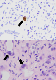
Adenovirus is a double-stranded DNA virus with more than 50 distinct serotypes. Though adenovirus infection is relatively common, adenoviral infection of the renal allograft is rare and also difficult to diagnose if viral inclusions are not present in the biopsy (see image above, black arrows indicating nuclear viral inclusion bodies). Based on a limited number of reported cases, patients typically present with fever, hematuria, dysuria, and acute kidney injury. Adenoviremia and viruria are present, as well as decoy cells similar to those seen with BK virus infection. Though it is possible that this patient may have had only ACR from the beginning (a close re-review of the 1st biopsy revealed no viral inclusions), it’s plausible that the adenovirus was responsible for the gross hematuria and thickened urothelium that were present from the start.

Adenoviral infection usually affects multiple organs and can be life threatening, though some patients are able to clear the virus and recover complete allograft function. After anti-viral treatment with intravenous cidofovir, our patient’s creatinine rose to 9 mg/dL. Unfortunately, she remains dialysis dependent and is undergoing re-evaluation for a second transplant.  This challenging case is a reminder that both rejection and infections must be considered when interstitial nephritis – and that one may be a decoy for the other!

Posted by Samira Farouk
Chief Fellow, Division of Nephrology
Icahn School of Medicine at Mount Sinai

Saturday, September 30, 2017

Nephrology Business Leadership University – A Fellow's Review.

Small Group Los Angeles Dialyspa NBLU 2017
In any academic fellowship, fellows receive excellent clinical training. However, the business aspect of practice often gets less attention. Before you know it you are scrambling to understand contracts, negotiate for a job, figure out billing/coding/reimbursement, understand market trends/growth, and figure out how to market your practice. Nephrology Business Leadership University or NBLU, aims to fill in this gap with a comprehensive 5-day course on understanding the business of Nephrology.

During our five days at NBLU, we learned the basics of insurance, 'red flags' of practices, the future of reimbursements, different practice environments (ie. academic, private, rural) and how to effectively interview/network among a variety of business related topics. Fellows were broken up into small groups and learned concepts in a variety of different teaching environments utilizing multiple interactive tools. In our small groups, we were assigned to a city and came up with a practice name as well as a mission statement. We were provided with an interactive map of our practice area and assessed growth rates to determine where we would open a clinic, which hospitals and dialysis units we planned to affiliate with. By far our favorite session was on billing/coding. This was an interactive session in which fellows were taught to 'think like a coder'; where we critiqued and assigned a level notes of other physicians.

By the end of NBLU, we were both feeling much more at ease about transitioning from fellowship to practice. I am ready to begin interviewing to find a practice that fits my needs. We highly recommend this conference to all freshly minted 2nd year Nephrology fellows interested in learning more about business and Nephrology.

However, the best part of NBLU is meeting and interacting with fellows from all over the country. We gained so much knowledge and now future friends and colleagues. How could you miss this opportunity? It is so much different than a brief meeting at Kidney Week or NKF as this is a 5 day immersion.

If you are interested in attending NBLU, pre-registration is now open for August of 2018, apply here

take some time to listen to the video testaments from this year’s attendees

Natasha Dave, Nephrology Fellow, Baylor Houston

Mona Shaban, Nephrology Fellow, University of North Carolina, Chapel Hill

From the Nate Hellman Unpublished Archive: Renal Ammonia Handling

Each day the average American creates 1 mEq/kg of acid which needs buffering. Most of the acid comes from protein. Our kidneys normally do all the buffering through 2 main systems – titratable and non-titratable acids Titratable acids (phosphates and sulfates) are relatively fixed in buffering capacity. Non-tritatable acids (ammonium – NH4+) does vary appropriately in patients having normally functioning kidneys. Thus, we adjust our buffering using ammonium.

If we cannot produce enough ammonium, then we develop a normal gap metabolic acidosis. Understanding the physiology of ammonium excretion helps us understand the possible problems.

  1. in the proximal tubular cells, glutamine converts to alpha ketoglutarate to produce NH3 (ammonia). Ammonia goes into the tubule and converts to NH4+. Thus, as patients lose proximal tubular cells they may lose buffering capacity because they cannot make enough ammonia.
  2. in the ascending limb of the loop of Henle, NH4+ goes into the intersitium by means of the sodium potassium 2 chloride cotransporter (this is the site which loop diuretics block). The NH4+ then converts back to NH3 and becomes part of the countercurrent multiplier system. Thus the medullary part of the kidney has very high concentrations of NH3.
  3. in the distal tubule, we acidify the urine. Here NH3 diffuses into the distal tubule, converts back to NH4+, creating the desired buffer effect.
Renal acidosis (normal gap) can occur for 3 major reasons.

  1. Distal RTA – inability to acidify the urine – cannot excrete the ingested acid if we cannot make mEq portions of NH4+ (which requires an acidified urine)
  2. Type IV RTA – this one is interesting. The problem in type IV RTA is primarily hyperkalemia (most commonly called hyporenin/hypoaldo). Increased potassium levels inhibit the enzyme glutaminase which is responsible for the production of ammonia. Thus if we correct the hyperkalemia, we usually resolve the acidosis.
  3. The acidosis of CKD – two problems for these patients – decreased production of ammonia and decreased concentration in the medulla. As CKD worsens, the countercurrent mechanism does not function as efficiently, decreasing the concentrations of solutes in the medulla. Thus, less ammonia is available to enter the distal tubule for buffering.

I hope these explanations make sense.

Friday, September 22, 2017

From the Nate Hellman Unpublished Archive: microRNAs and the Kidney

MicroRNAs: one of the sexiest topics in the science community these days! Pick up a copy of Science, Nature, or Cell and you can usually find something to do with microRNAs within. What do these have to do with the kidney? Well, it's still a little premature, but there are certainly microRNAs present in the kidney, and recent studies have demonstrated that specific microRNAs are upregulated in conditions such as polycystic kidney disease and renal cell carcinomas, for instance.

How microRNAs work: microRNAs are single-stranded RNA molecules of between 21-23 nucleotides in length which are partially complementary to regions in multiple mRNAs. Once they bind to these mRNAs, the microRNAs either inhibit translation or completely degrade their target RNAs. A specific enzymatic machinery--comprised of the proteins Dicer and the RISC complex--is responsible for inhibiting mRNAs via microRNAs.

In essence, the power of microRNAs are that a single microRNA can regulate the expression of multiple genes working in parallel to achieve a similar biologic effect. This technology is of particular use to the pharmaceutical industry: one can envision targeting a particular microRNA which inhibit several pathways to prevent a disease process, such as atherosclerosis, renal fibrosis, or cyst formation to think of a few possibilities. The field is still very new. I wouldn't be surprised if a future Nobel Prize came out of this work. Three scientists (Drs. Ruvkun, Baulcombe, and Ambros) working on microRNAs recently won the 2008 Lasker Prize--considered by many to be the "precursor" prize to the Nobel.

Thursday, September 21, 2017

Nephrology Fellows: last call to join an online focus group on palliative care


Calling all Nephrology fellows!

Duke University School of Medicine is seeking to understand barriers and facilitators to palliative care conversations between nephrology fellows and their patients. The study, funded by the Agency for Healthcare Research and Quality, is led by Jennifer St. Clair Russell, PhD, MSEd, MCHES, a social and behavioral scientist in the Division of General Internal Medicine with over 11 years of experience in nephrology.

 If you are eligible to participate in our online focus group (on September 28), you will be compensated for your study participation. It is estimated that the online focus group will only take approximately 120 minutes.

We would be honored to have you participate in this exciting research!

To learn more or complete the eligibility questionnaire, please contact Nikita Shah at Nikita.Shah@duke.edu.

Monday, September 18, 2017

A Primer on Primary Hyperoxaluria

Primary hyperoxalurias are a group of autosomal recessive disorders characterized by excess hepatic oxalate production and reduced kidney excretion.

They are classified into three types based on their enzymatic defect in glyoxylate metabolism
  • Type 1 (80%): alanine glyoxylate aminotransferase (AGT) 
  • Type 2 (10%): glyoxylate reductase/hydroxypyruvate reductase (GRHPR) 
  • Type 3 (10%): mitochondrial 4-hydroxy-2-oxoglutarate aldolase enzyme (HOGA1)
Variable in presentation and severity, its true prevalence is likely underestimated. While it can occur at any age, the median age of onset is 5.5 years. In Europe, it has an estimated prevalence of 1-3 cases per 1 million, incidence of 1 case per 120,000 live births and account for 1-2% of pediatric ESKD Unfortunately 20-50% of patients have advanced CKD at diagnosis and 10% are diagnosed only after disease recurrence following transplantation.

Excreted almost entirely by the kidney, oxalate can cause kidney tubular toxicity, nephrocalcinosis and obstruction with superimposed infection. In type 1 primary hyperoxaluria as eGFR falls below 30, plasma oxalate rises resulting in oxalate deposition in various organs including the kidney, heart, joints, retina, skin, bone marrow, heart and CNS.

Given its rarity, clinical suspicion is required for diagnosis. Definitive diagnosis requires genetic testing. Clinically it presents with urolithiasis (greater than 95% calcium oxalate monohydrate), recurrent UTIs, metabolic acidosis, acute kidney failure or oxalate crystals on kidney biopsy. Labs can be notable for persistently elevated urine oxalate, serum L-glycerate or serum oxalate. For persons with a family history, prenatal diagnosis can be obtained through genetic screening in the first trimester.

Conservative treatment strategies should be employed early and include aggressive hydration and potassium citrate to inhibit crystallization. Pyridoxine supplementation can be used for primary hyperoxaluria type 1 Gly170Arg and Phe152Ile genotypes. Intestinal oxalate has limited effects on disease progression as oxalate is largely produced endogenously and studies using oxalate-metabolizing probiotics like Oxalobacter formigenes have been disappointing.

In primary hyperoxaluria type 1 conventional HD and PD are ineffective at removing sufficient levels of oxalate for patients with ESKD due to ongoing liver production. While the ultimate management is transplantation, dialysis may be required as a temporary therapy to reduce plasma oxalate levels. Strategies include short daily sessions of high-flux dialysis, nocturnal dialysis, or combinations of hemodialysis and nocturnal peritoneal dialysis.

Isolated liver transplantation corrects the enzyme defect and can be used in early stages of kidney disease. Isolated kidney transplantation can reduced plasma oxalate, but disease recurrence often leads to poor graft survival. Dual liver-kidney transplantation is thus commonly performed for patients with CKD stage 4. Limited data exists for organ transplantation in primary hyperoxaluria type 2 and type 3 has not yet been associated with ESKD.

Sapna Shah, MD
Nephrology Fellow
Mount Sinai, New York

Tuesday, September 12, 2017

Prune Belly Syndrome and Kidney Disease


Prune Belly Syndrome (PBS) is a very rare (3.8/100,000 live births) congenital disease that predominantly affects males.

PBS is characterized by a triad of
  • Anterior abdominal wall muscle deficiency 
  • Urinary tract abnormalities
  • Bilateral cryptorchidism. 
Termed prune belly due to the wrinkled appearance of the abdominal wall at birth, it is a multisystem disease that can affect the genitourinary tract, cardiopulmonary, GI and musculoskeletal systems. Some studies and the inheritance pattern suggest that the disease is due to a recessive X-linked defect.  However, other studies have demonstrated a deletion in the the hepatocyte nuclear factor 1 beta (HFN1B) gene on chromosome 17q12 resulting in defective mesenchymal development, thus suggesting autosomal dominant inheritance. A clear genetic basis for PBS has not yet been established.

As per Seidal et al., nearly one-half of patients surviving infancy will develop CKD and nearly one third will develop ESRD and require RRT. This is thought to occur largely due to incomplete nephron differentiation, dilation of tubules, interstitial fibrosis and eventual glomerulosclerosis. Clinically PBS can be associated with reflux nephropathy and recurrent pyelonephritis.

Often diagnosed at birth or in early childhood, PBS can be diagnosed by antenatal ultrasound between weeks 20-30 with features including hydroureteronephrosis with or without echodense kidney parenchyma, oligohydramnios, hypoplastic lungs and bulging abdomen.

Therapy in childhood remains controversial, but can require early surgery for urine drainage in order to prevent recurrent infections or antibiotic prophylaxis. Maintenance CAPD and HD are equally effective in the management of ESKD and kidney transplantation has been successfully performed with excellent long-term prognosis.

Sapna Shah, MD
@NephShah
2nd Year Nephrology Fellow
Icahn School of Medicine, Mount Sinai, New York


Monday, September 11, 2017

Attn Fellows: Participate in an Online Focus Group on Palliative Care

Calling all nephrolgoy fellows!

Duke University School of Medicine is seeking to understand barriers and facilitators to palliative care conversations between nephrology fellows and their patients. The study, funded by the Agency for Healthcare Research and Quality, is led by Jennifer St. Clair Russell, PhD, MSEd, MCHES, a social and behavioral scientist in the Division of General Internal Medicine with over 11 years of experience in nephrology.

If you are eligible to participate in one of our two online focus groups (either September 17 or September 28, 2017), you will be compensated for your study participation. It is estimated that the online focus group will only take approximately 120 minutes.

We would be honored to have you participate in this exciting research!

To learn more or complete the eligibility questionnaire, please contact Nikita Shah at Nikita.Shah@duke.edu

Saturday, September 9, 2017

Origins of Renal Physiology at MDIBL Continues to Inspire Nephrology Fellows

2017 MDIBL Origins of Renal Physiology National Renal Fellows Course
I first discovered the Origins of Renal Physiology course on Renal Fellow Network as a medicine intern, and ever since that time, dreamt of going to the course as a renal fellow. Nate’s picture in front of the “Kidney Shed” was seared in my mind. The dream finally came true two weeks ago when I set foot on the Mount Desert Island Biological Lab (MDIBL) in Coastal Maine, and the first place I visited was indeed the Kidney Shed.

In his opening address to the fellows attending the course, Dr. Ziedel rightly said “Physiology is the pride of Nephrology”. He went on to mention the names of several luminaries who have worked at the MDIBL leading to many important discoveries. The lab has been a cornerstone of nephrology research and has been a home for the work of Homer Smith, E.K Marshall, David Evans, Franklin Epstein, and many others who have spent years working on and elucidating the fundamental concepts on kidney physiology.

In modern day practice of nephrology, we are less exposed to these fundamentals of kidney physiology that initially piqued our interest in nephrology. The Origins course is one of the many efforts made by the renal community to reconnect nephrolgoy fellows to physiology. It’s a very unique course which gives the fellows an opportunity to rub elbows with senior and distinguished renal physiologists from esteemed institutions and work with them on experiments to understand the different aspects of kidney biology.

The course consisted of 7 modules, each focusing on a functional unit of the nephron- the glomerulus, proximal tubule, thick ascending limb, distal tubule, acid-base, water homeostasis, and chloride secretion. We studied animals like zebrafish, turtles, and toad bladders. We also explored how ENaC and NKCC channels work. We delved into discussion about morpholinos and so much more. Over the duration of the course, we developed a new-found appreciation for the beauty and wonder of exploring how the kidney regulates homeostasis. We also got a sense for the profound impact MDIBL has had on the field of nephrology and physiology.

As MDIBL is in such close proximity to Acadia National Park- one of the most beautiful national parks in the world, nature is inherently embedded in the process of learning here. Most days started off with a refreshing early morning short hike which energized us to spend the entire day in the lab. On alternate days, amazing outdoor activities were planned for us after presentations of the earlier day’s work. My week at the MDIBL during this course was very memorable.

I was honored to be working closely with great mentors and met nephrology fellows from all over the US and abroad, shared interests and future aspirations and found new friends. 

All in all, an incredible experience! I would wholeheartedly endorse this conference for future fellows.

Manasi Bapat
@manasib33
2nd year Nephrology Fellow
Mount Sinai Hospital, NY

Tuesday, September 5, 2017

September Wash U Nephrology Webisode available - an unusual case of AKI!

Another great episode on the road, as I packed up my gear and headed to Rush University Medical Center in Chicago, IL.  Discussants joining me for this case are Dr. Roger Rodby, Dr. Pravir Baxi, and Dr. David Cimbaluk.  More complicated histology in a very unusual case of AKI:

"73 year old Caucasian female with hypertension, remote history of breast cancer, and NSAID use, presents with incidental AKI"


Tuesday, August 22, 2017

A few months ago, we posted about an acid-base primer that was being offered free for fellows in the US.

During the 5-day period from Wednesday, August 23 - Sunday, August 27, you can get a free copy of this ebook (kindle) edition of The Painless Guide to Mastering Clinical Acid-Base. Simply go to Amazon, search the title of Amazon ID number (B06XRM56TY), or click here, and you will find the ebook priced at $0.00 during those 5 days. Once you "purchase" the book, it will remain permanently in your Amazon library and you will be able to load it onto any device that has a Kindle app linked to your account. Feel free to notify students, residents, nurses or any others who might benefit. There is no limit to the number of persons who can get the download.

Posted by Ben Abelow