Showing posts with label Ailish Nimmo. Show all posts

Wednesday, March 14, 2018

The Uncertainty of Monoclonal Gammopathies

I spend a lot of time discussing patients with renal disease and monoclonal gammopathies with my haematology colleagues, trying to figure out what is of ‘renal significance’ and ‘undetermined significance’. One of those discussions recently was around a normally well 70 year old lady. She presented with several pre-renal insults including a febrile illness, relative hypotension and NSAID use for joint pains. Her renal function rapidly deteriorated and she became oligoanuric requiring haemodialysis.  

Image result for immunotactoid GNAt a routine medical 4 months previously she was normotensive with normal renal function and no proteinuria.  Investigations during this admission revealed nephrotic range proteinuria and an IgG kappa paraprotein at 1g/L with a normal free light chain ratio.  Autoantibodies and cryoglobulins were negative. A renal biopsy showed diffuse endocapillary glomerulonephritis with immunoglobulin pseudo-thrombi in the capillaries and negative Congo Red staining.  Staining was positive for all immunoglobulins and C3. Electron microscopy showed organised sub-endothelial deposits of hollow microtubules of 40-50nm diameter in keeping with immunotactoid glomerulopathy (see image). A bone marrow showed no significant dysplasia, no light chain restriction and no evidence of myeloma. She was treated with high dose steroids and initially plasma exchange whilst results were returned.  Her renal function improved over the following week and she did not require further specific therapy given the absence of overt lymphoproliferative or autoimmune disease.
It is not surprising that we pick up a lot of monoclonal gammopathies in patients with deteriorating renal function or proteinuria as MGUS affects 3% of patients over the age of 50. Recent evidence suggests that MGUS progresses to myeloma or other plasma cell or lymphoproliferative disorders in 10% of patients at 10 years, and higher than this in those with an abnormal serum free light chain ratio or IgM paraprotein.
Monoclonal immunoglobulins or immunoglobulin-derived fragments can be deposited in the kidney in a number of patterns (previous RFN post from Nate):
  • Crystalline deposits: cast nephropathy in myeloma
  • Organised fibrillar deposits: AL amyloidosis (Congo Red stain positive)
  • Organised microtubular pattern: immunotactoid GN or type I cryoglobulinaemia.  Fibrillary GN is from polyclonal IgG deposition (not usually associated with a paraprotein) and on EM has slightly smaller fibrils (approx. 20nm in diameter) than immuntactoid GN.
  • Disorganised granular deposits as in LCDD and HCDD 

Immunotactoid GN usually presents in a more insidious pattern with hypertension, haematuria and proteinuria rather than acutely as in this case.  The straight, hollow tubules are over 30nm in diameter and are usually composed of crystallised monoclonal IgG. The diagnosis can be missed when EM is not performed.  It is usually secondary to lymphoproliferative diseases, typically CLL or B cell non-Hodgkin’s lymphoma but also MGUS. Treatment is based on the underlying disease, and in cases as this vigilance for progression is vital.  


Post by Ailish Nimmo

Friday, December 29, 2017

The ELITE & the Rest in Kidney Transplantation

Image result for cocktail party
The ELITE-Symphony study enshrined standard triple therapy immunosuppression of tacrolimus, mycophenolic acid and prednisolone, for renal transplant recipients. We get familiar with these agents relatively quickly in our renal training, but what about the other less common agents that are used?  Why do we not use them as first line, when should we consider them and what side effects do we need to be aware of?
Sirolimus and everolimus are mTOR inhibitors (see previous RFN post), and prevent cell cycle progression and lymphocyte proliferation.  Their anti-proliferative effects mean they have a role in conditions such as tuberous sclerosis, psoriasis and certain cancers.  The antiproliferative and antiangiogenic effects pose issues post-surgery however with poor wound healing and lymphocele formation meaning they should not be used for around 6 months post-transplant.  Other side effects include pneumonitis, hyperlipidaemia, bone marrow suppression, thrombotic microangiopathy and proteinuria due to FSGS-type lesions. Alongside this relatively long list of side effects, the main concern with sirolimus is related to mortality. A meta-analysis of 21 studies with a total of 5876 patients showed sirolimus was  associated with increased mortality post-transplant, mainly from cardiovascular and infectious complications (adjusted hazard ratio 1.43, 95% CI 1.21-1.71). So why do some patients end up on sirolimus?  The same meta-analysis showed that sirolimus is associated with a 40% reduced risk of malignancy, particularly in non-melanoma skin cancers. The most common reasons for switching to mTORi are malignancy, often recurrent skin cancers, and for calcineurin-inhibitor (CNI) induced injury (although if eGFR<40mls/min or overt proteinuria outcomes are likely to be poor or no better with a switch). Moreover, as seen in ELITE-Symphony and other, acute rejection rates and patient dropouts are consistently higher with mTORi compared to CNIs.
Image result for belatacept mechanismAnother agent we may be hearing more about in the future is belatacept: a co-stimulation pathway blocker. It is attractive as it is designed to replace the CNI in the treatment regime. It binds to CD80 and CD86 on antigen-presenting cells, thereby blocking CD28 mediated activation of T Cells. It is administered as an IV infusion every 4-6 weeks alongside steroid and an anti-metabolite.
The BENEFIT and BENEFIT-EXT (extended criteria donors) trials compared high and low intensity belatacept regimes with cyclosporine in non-sensitised recipients undergoing DBD or living donor renal transplants. Over 7 years of follow-up, they found improved patient and graft survival with belatacept in BENEFIT and better measured GFR and reduced formation of DSA with belatacept in both studies. There was an increased incidence of acute rejection in BENEFIT although this obviously didn’t translate into inferior outcomes. There were also initial concerns regarding a small increase in cases of PTLD in EBV seronegative recipients although longer term data was reassuring that the overall risk of this was very low.
A potential benefit of belatacept is it’s a directly observed therapy, so compliance will be known. Although it is expensive, if patients experience improved graft outcomes then any longer term savings need to be considered. However what about patients who are highly sensitised? Many patients we want to avoid CNIs in are sensitised but here is a dearth of data in using belatacept in this circumstance. Moreover, the studies compared belatacept to cyclosporine, not tacrolimus, the current standard of care. Currently belatacept is not available for new patients due to supply issues, another issue likely preventing its more widespread use.  
Post by Ailish Nimmo

Friday, June 30, 2017

An Under Recognised Cause of Metabolic Acidosis

The MUDPALES mnemonic for raised anion gap acidosis was drilled into me from medical school.  However recently after working through each category I became stumped when nothing ticked the box to identify the cause. 
The lady I had been asked to see was in her 80s and had fractured her hip.  On admission she had normal renal function and acid base status.  Post-operatively she was started on regular analgesia including paracetamol (acetaminophen) and developed a Staphlococcus Aureus wound cellulitis treated with flucloxacillin.  Over the following 2 weeks she developed a raised anion gap acidosis and positive urinary anion gap.  Renal function, lactate and ketones were normal.    
A cause of metabolic acidosis not in MUDPILES is pyroglutamic acidosis.  Pyroglutamic acid (also called 5-oxoproline) is a by-product in the gamma-glutamyl cycle.  This pathway is involved in the synthesis of glutathione, and is shown above from a recent paper.
Glutathione provides negative feedback on the cycle by inhibiting the enzyme gamma-glutamyl-cysteine synthase.  An acquired deficiency in glutathione, as with alcohol or paracetamol, results in loss of this negative feedback and increased production of 5-oxoproline leading to a metabolic acidosis.  Other drugs affect the cycle at different points including flucloxacillin which inhibits 5-oxoprolinase similarly resulting in build-up of 5-oxoproline. 
Urine amino acid screens show high levels of 5-oxoproline, though this test is not always available.  Our patient improved with withdrawal of paracetamol and flucloxacillin and oral bicarbonate which was stopped after a week.  Some cases have been treated with N-acetylcysteine to replenish glutathione levels. 
I think this is probably an under-recognised cause of metabolic acidosis with many of the risk factors being prevalent in our inpatients (poor nutrition, chronic alcohol use, renal failure, infection, paracetamol use).  A different mnemonic for causes of raised anion gap metabolic acidosis may be helpful like GOLD MARK:
·         G – glycols (ethylene and propylene)
·         O – 5-oxoproline (pyroglutamic acid)
·         L – L-lactate
·         D – D-lactate
·         M – methanol, ethanol
·         A – aspirin/salicylates
·         R – renal failure
K – ketones 

Post by Ailish Nimmo

Friday, April 14, 2017

Jaundice and Renal Failure

In patients with combined renal and liver dysfunction it is important to differentiate between renal impairment that is secondary to liver disease i.e. hepatorenal syndrome, and conditions where an insult directly affects both the liver and kidney.  
I recently saw a 20-something year old student who was a regular kayaker in local rivers.  He had a 5 day history of myalgia, fever and headache with no vomiting or diarrhoea.  He took paracetamol (acetaminophen) and ibuprofen within recommended limits for his symptoms.  In the 24 hours prior to his admission he developed jaundice and noticed decreased urinary output. On arrival to hospital he was jaundiced but examination was otherwise unremarkable.  There were no signs of chronic liver disease and the liver was not palpable.  He was hemodynamically stable.  Blood tests showed a raised (conjugated) bilirubin at 453umol/L (25 mg/dL), ALT within normal limits and mildly elevated ALP at 162U/L.  He had an acute kidney injury with creatinine 427umol/L (4.83 mg/dL), potassium 3.4mmol/L.  He was anemic and thrombocytopenic with a neutrophilia, and blood film showed toxic granulation, with no red cell fragments.  An ultrasound of his liver was unremarkable and his kidneys were at the upper limit of normal size with no hydronephrosis. For 36 hours he was anuric despite fluid resuscitation, and required a short period of CRRT.  After this time he began passing urine and his creatinine spontaneously fell, although bilirubin remained elevated.  He was treated empirically with ceftriaxone and doxycycline for leptospirosis, and six days later urine PCR and serum IgM came back positive for leptospirosis.  
Flores-Figure-2-comp
Photo from ACG Case Reports Journal


Leptosporosis is a zoonosis usually transmitted to humans through rodent urine in water.  The majority of cases cause a self-limiting febrile illness, but in its severe form - Weil’s syndrome – patients can develop jaundice, renal failure, pulmonary haemorrhage and aseptic meningitis.  Renal impairment in leptospirosis can be secondary to a number of different mechanisms including sepsis/critical illness, a direct nephrotoxic effect to the renal tubule by the leptospira toxin, tubulointerstitial nephritis, rhabdomyolysis and hyperbilirubinemia.  Patients often maintain good urine output despite renal injury, and tubular dysfunction means most patients are hypo- or normokalemic even if oliguric.  Another characteristic finding is enlarged kidneys with normal parenchymal echogenicity on ultrasound.  
Included in our initial differential was that his renal impairment was secondary to hyperbilirubinemia.  Bilirubin causes renal impairment again through a range of mechanisms including direct toxicity to the nephron, bile acid casts causing tubular obstruction, and hypoperfusion from vasodilation.  Renal biopsy can show pigmented bile casts within the tubules and tubular hypertrophy (see image).  Electron microscopy can show bile acid accumulation within lysosomes and dilated mitochondrial cristae.  In those with normal baseline renal function and short-lived hyperbilirubinemia changes are often mild and reversible, but irreversible damage can occur in those with underlying renal impairment.  
Early clues for leptospirosis in this case include hypokalemia despite being oligoanuric, enlarged kidneys on ultrasound and of course his social history.  


Post by Ailish Nimmo

Tuesday, February 21, 2017

Spontaneous Tumor Lysis Syndrome

A 70 year old Ghanaian man was recently admitted under our care.  He had been diagnosed with aggressive myelodysplasia 2 months previously after presenting with fatigue and abnormal blood results (WBC 50.3, platelets 130 and LDH 928 at the time of diagnosis).  A plan was made for palliative chemotherapy. One month after his diagnosis he developed a large pericardial effusion and had 1L of haemorrhagic fluid drained.  At this point his creatinine was 200 umol/L (2.26 mg/dL).  Routine and TB culture of the fluid was negative, as was cytology and immunophenotyping.
Image result for swissnephro uric acid
Two weeks after this admission he represented with abdominal pain.  A CT showed bilateral renal and bladder calculi without obstruction.  He was oliguric with a creatinine of 577 umol/L (6.5 mg/dL) rising to 709 umol/L (8 mg/dL) over the next 12 hours.  His uric acid level was 18.0 mg/dL, which had not been checked previously.  Phosphate was 1.86 mmol/L (5.75 mg/dL), Ca 2.1 mmol/L (8.4 mg/dL) and K 4.6 mmol/L.
Our diagnosis was of a spontaneous tumour lysis syndrome (TLS; see previous RFN posts here & here).  Nucleic acids released from tumour cell lysis are broken down into xanthine and then uric acid by xanthine oxidase.  Renal failure is caused by uric acid precipitating in renal tubules causing a mechanical obstruction and inflammatory reaction.  While TLS is typically seen following initiation of chemotherapy causing a rapid breakdown of cancer cells, a spontaneous form has been described in acute leukaemia and NHL.  Our patient was at high risk of converting into AML but had no rise in peripheral blasts to suggest this.
Interestingly, spontaneous tumour lysis syndrome is associated with hyperuricemia but often without the hyperphosphatemia (and hyperkalemia) seen in the classical form of the disease– thought to be because the released phosphorus is quickly used up in the generation of new tumour cells.  This would fit with our patients results.
Our patient was commenced on dialysis which gave reductions in uric acid levels of 50% per treatment, but they quickly rebounded.  He was no longer fit for treatment of his myelodysplasia making longer term management more difficult.  Given his African ethnicity, we checked his glucose-6-phosphatase levels, which were normal, before he received rasburicase (recombinant urate oxidase). Rasburicase reduces uric acid levels by converting it into allantoin.  It may cause severe oxidative hemolysis if glucose-6-phosphatase deficient. Uric acid fell to undetectable levels following this however he had an ongoing dialysis requirement (note that rasburicase retains in vitro activity in the blood bottle so sample should ideally go on ice).  Allopurinol as a longer term medication to reduce uric acid formation may be useful, but may not manage to suppress formation sufficiently.  
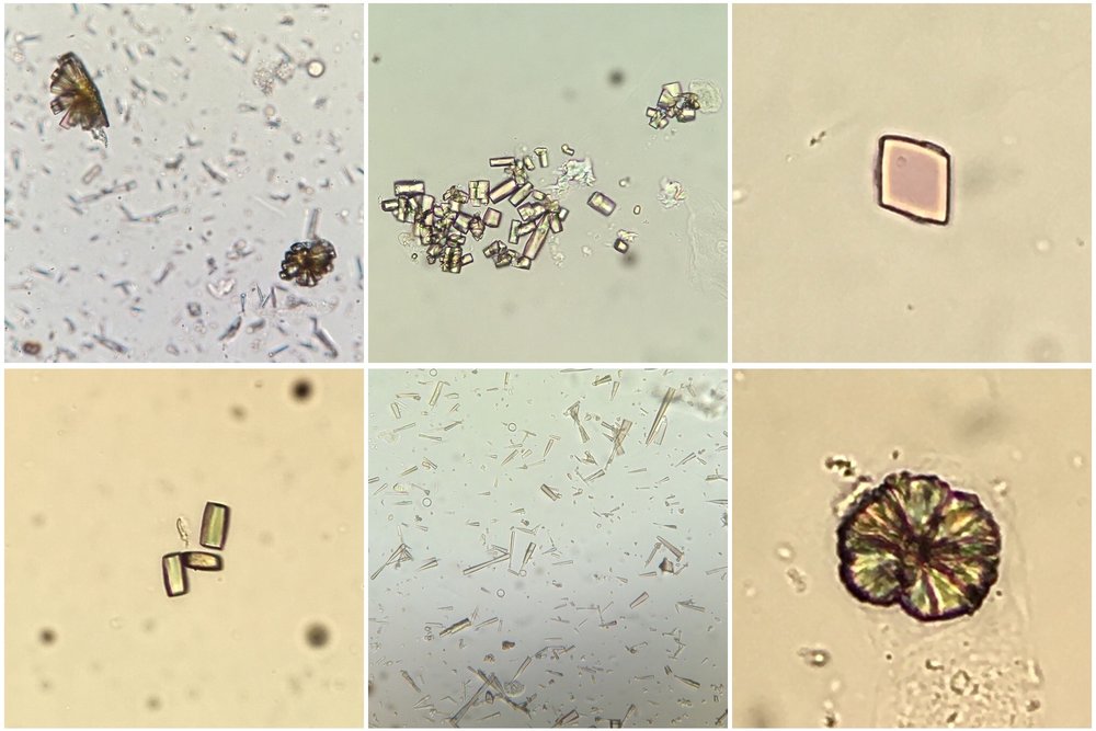
In addition to tumour lysis syndrome, acute urate nephropathy can be caused by other states of tissue catabolism such as seizures, in primary overproduction of uric acid or in cases of reduced urate reabsorption in the proximal tubule. Urinalysis can show uric acid crystals (birefringent with polarisation; see image) or can be normal (as in our patient) perhaps due to a lack of output from obstructed tubules. 
This case raised several points to me. Was his pericardial effusion also caused by a urate infiltration?  No clear cause was ever identified at the time and he did not appear ‘uremic’ despite his renal dysfunction.  Could any of this have been prevented if treatment for his hyperuricemia had been commenced earlier?  I also learned:
  • The nuances of spontaneous tumor lysis syndrome (often phosphate & K not hugely elevated).
  • Rasburicase is contraindicated if glucose-6-phosphatase deficient (approximately 20% of Africans).
  • The ‘undetectable’ result of urate after rasburicase administration appears to be due to in vitro activity of the drug in the blood bottle.

Image thanks to Florian Buchkremer @swissnephro

Post by Ailish Nimmo

Wednesday, May 4, 2016

Emphysematous Pyelonephritis


http://www.indianjnephrol.org/articles/2013/23/2/images/IndianJNephrol_2013_23_2_119_109418_f6.jpgI was asked to see 74 year old man with an acute on chronic kidney injury.  He had 2 days of generalised lower abdominal pain and vomiting but no urinary symptoms or fever.  His past history included advanced CKD, benign prostatic hypertrophy and a slow growing renal cell tumour under radiological surveillance.  His vital signs were normal and he had mildly raised inflammatory markers.  I ordered a CT KUB to exclude obstruction (it was a weekend and was no ultrasound service in the hospital).  To my surprise this came back as showing emphysematous pyelitis.  Interestingly there had been a hiss of air as he was catheterised for fluid balance monitoring – a fact I had dismissed at the time!


Emphysematous UTIs are gas forming infections of the urinary tract and can manifest as cystitis (gas within the bladder wall), pyelitis (gas within the collecting system) or pyelonephritis (gas within renal parenchyma or perinephric tissues).  It is a relatively rare condition and there is a dearth of literature describing incidence.  Diabetes and urinary tract obstruction are major risk factors, present in around 80% and 20% of patients respectively. Causative organisms are most commonly E. Coli and Klebsiella pneumoniae, with Candida being involved less frequently. Presentation is usually similar to acute severe pyelonephritis with fever, flank pain and vomiting. 50% of patients have an associated bacteraemia. Diagnosis is usually made by CT which shows the extent of gas within the urinary tract and any obstruction.
Treatment depends on the extent of infection.  It ranges from parenteral antibiotics alone for patients where gas is limited to the collecting system with no obstruction, to percutaneous drainage of purulent material and antibiotics if there is abscess formation or extension of gas into the perinephric space, to nephrectomy in patients with diffuse gas and extensive renal destruction.

In the above case, the urine sample was initially reported as ‘no significant bacteriuria’ but subsequently grew a resistant E. Coli >1,000 - <10,000 cfu/ml.  Urology felt that surgical intervention was not required as the renal parenchyma was not involved and he had no abscess formation.  The patient completed 2 weeks of ertapenem and his renal function returned to baseline.
I took several learning points away from this case:
  • As someone who spends a lot of time signing off patients’ results, I realise that ‘no significant bacteriuria’ is not the same as ‘no growth’, and in this case the difference was substantial.  The wording of how we report things and how we interpret that is crucial.
  • A high index of suspicion is required to diagnose emphysematous UTIs and the most appropriate imaging modality should be considered.  Ultrasound is generally the first line investigation for urinary obstruction in patients with acute kidney injury or febrile urinary tract infection due to high sensitivity for hydronephrosis, lack of ionising radiation and lower cost than CT. Ultrasound appearances in emphysematous UTIs can be difficult to interpret however: gas, calculi and calcifications are hard to distinguish and there is often variability in how they are reported. CT is able to precisely localise the presence of gas within the urinary tract and determine whether there is involvement of the renal parenchyma and perinephric tissues.  It can also identify any concomitant pathology or alternate diagnosis e.g. renal calculi.  CT is therefore preferable for diagnosis and subsequent severity staging.
  • Pneumaturia has been described as a presenting feature of emphysematous UTI.  Other causes include vesicovaginal or vesicoenteric fistulae, renal tumour infarction and recent instrumentation.  The unexpected air hiss when catheterising this gentleman was a warning of a more serious pathology and should prompt further investigation.

While the outcome of emphysematous pyelitis is better than that of pyelonephritis (which has a mortality of 18-70% depending on extent of involvement), it is still not a condition to be taken lightly. 
Post by Ailish Nimmo, Royal Infirmary of Edinburgh