Showing posts with label cancer. Show all posts

Sunday, June 21, 2015

Renal Effects of Anti-Cancer Medications

With the proliferation of new therapies for cancer including novel targeted therapies, it is difficult to keep up with the potential renal problems related to these medications. Thankfully, Nature Reviews Nephrology have published a comprehensive review of the nephrotoxic effects of targeted anticancer therapies that could serve as a reference for practicing nephrologists. As is pointed out in the review, this is a rapidly evolving field and novel toxicities are being recognized with real world use that may not have been seen in the initial trials..

Of particular interest was the fact that many of these agents cause severe electrolyte abnormalities. For example, approximately 17% of individuals receiving cetuximab (an EGFR inhibitor used in the treatment of some lung cancers) develop hypomagnesemia. It turns out that TRPM6, a DCT  magnesium channel, it regulated by EGF which is highly expressed in that area of the nephron and cetuximab therapy leads to inhibition of Mg reabsorption and subsequent hypomagnesemia (along with hypocalcemia and hypokalemia).

The figure below shows a list of the commonest adverse effects of chemotherapeutic agents. Click on the picture for a larger image:


Sunday, February 1, 2015

ESAs in Patients with CKD and Cancer: Is the Risk Worth the Benefit?

Anemia is a common manifestation of CKD. Currently, there are no guidelines for nephrologists regarding erythropoietin stimulating agents (ESAs or Epo or Darbe) use in patients with CKD with previous or active malignancy. Recently an excellent review of this topic was published in Kidney International by Hazzan et al. Let’s go over some of the key points.

Erythropoietin Biology and Relevance in Malignancy
  • Besides stimulating erythropoiesis, Epo has been shown to have both anti-apoptotic and pro-proliferative actions in endothelial cells, brain +/- spinal cord, kidney and heart. Furthermore, Epo has also been shown to promote angiogenesis in endothelial cells. These non-erythroid functions of Epo are not fully understood.
  • Epo mediated angiogenesis appears to be physiologic and driven by hypoxia but may play pathological role in proliferative diabetic retinopathy.
  • Because angiogenesis is important for tumor survival and progression, it is important to know if cancer cells express the EPo receptor (EpoR). Initial evaluations for the EpoR on cancer cells, tested either for the Epo- receptor antibody (the antibodies used were non-specific) or mRNA transcripts (which were potentially contaminated by other cell types from blood or stromal tissue) and were less informative. Recent development of first specific antibody to the EpoR (A82) with both positive and negative controls will allow more rigorous testing for the EpoR protein on cancer cells. Swift et al. studied 66 cell lines and found either no or very low levels of the EpoR protein. Although current data does not show strong evidence of presence of functional EpoR on cancer cells, we need more evidence to draw a definite conclusion.
  • In absence of EpoR, Epo induced supra-physiologic Hb levels would increase oxygen delivery to cancer cells and potentially stimulate proliferation.
  • Also, hypothetically, binding of Epo to EpoR expressed on activated macrophages can suppress NF-kB activation and proinflammatory genes, resulting in an immunosuppressive effect. 
Nephrology Literature on ESA usage in Patients with CKD and Cancer
  • The TREAT trial was a landmark study in the field of nephrology which was published in 2009 in the NEJM. The TREAT (and CHOIR) trials changed how we treated anemia. In this study, more than 4000 diabetic CKD patients with anemia were randomized to either higher hemoglobin (Hb) target (13 g/dl) with darbepoetin or lower hb target (9 g/dl) in placebo arm. Surprisingly, there was a trend towards increased risk of death due to cancer in the Epo group (darbepoetin alfa group 39 deaths, placebo group 25 deaths, P=0.08). Also, in patients with a previous history of cancer, there was increased mortality due to malignancy in Epo group (darbepoeitin alpha 14/188 deaths, Control 1/160 deaths, P=0.002). These results, for the first time, raised concern regarding possible association of Epo with cancer.
  • However, a year later in 2010 Japanese study in CKD stages 4 and 5, failed to show an increased incidence of cancer with Epo. But this study targeted lower Hb (10.1 g/dl) and had a short follow up period.
  • Seliger et al found that Epo increased the risk of stroke, only in CKD patients with diagnosis of cancer. But cancer group received higher initial ESA dose even though the pre-ESA Hb was similar in both groups.
Oncology Literature on ESA use in Patients with CKD and Cancer

Head and neck cancer trials had used Epo to increase tumor oxygenation in an effort to increase efficacy of radiotherapy.
  • ENHANCE TRIAL 2003 was conducted in head and neck cancer patients given ESAs while under-going only radiotherapy (no chemotherapy). Surprisingly, locoregional progression-free survival was found to be poorer with epoetin (where patients were treated to Hb 14–15 g/dl) than with placebo.
  • Similar results were found by DANISH RCT which reported 10% difference in 3-year local/regional control in favor of the control group (P=0.01) compared to darbepoetin group 
Meta-Analysis of Outcomes of Mortality with ESAs
  • Cochrane database analysis- Dec 2012 , found strong evidence that ESAs increased mortality during the active study periods (death occurring up to 30 days after active study protocol) (hazard ratio 1.17; 95% CI 1.06–1.29), and borderline evidence that ESAs decreased overall survival (hazard ratio 1.05; 95% CI 1.00–1.11). The increase in mortality risk was seen in studies where patients had Hb higher than 12 g/dl before Epo treatment. 
  • Mortality risk was higher in patients who received Epo without concurrent chemotherapy but these trends in patients receiving Epo and concurrent chemotherapy are not clear.
  • There is insufficient evidence to know if the risk is dependent on the cancer type. 
Clinical Implications and Recommendations for ESA use in Patients with CKD and Cancer 
1. The nondialysis CKD/ESRD patient with current cancer:
  • Only FDA indication for ESA treatment is for anemia caused by current myelosuppressive chemotherapy; there is no indication for patients with cancer not receiving chemotherapy.
  • If acute severe, symptomatic anemia is present, then blood transfusion is the treatment of choice. 
  • Suggest generally limiting the Hb target to an upper level of 10 g/dl to prevent risk of stroke and mortality with higher Hb targets.
  • For the occasional patients who still have anemia-related symptoms, a slightly higher Hb target may be considered. 
  • Intravenous iron may be given to minimize total ESA dose exposure.
  • The FDA and some oncology guidelines recommend against the use of ESAs if chemotherapy treatment is with curative intent.
  • Hazzan et al feel that ESA treatment is probably reasonable for the advanced non-dialysis CKD/ESRD patient receiving chemotherapy with curative intent but with an upper Hb target of only 10 g/dl. However patient counselling of the risk and benefits is mandatory before Epo use. 
2. The nondialysis CKD/ESRD patient with a previous history of cancer:
  • Discuss with patient`s oncologist if the cancer is cured. Ask about risk of recurrence and the risk for other tumors related to the primary malignancy or its treatment.
  • For up to 5 years after potential cure, treat with ESAs as if active cancer was present, maintaining an upper limit of Hb of 10 g/dl.
  • Even after confirmed cure with very low risk for recurrence, make efforts to reduce ESA dose exposure by ruling out other treatable causes of anemia. (Remember TREAT trial) 
3. The nondialysis CKD/ESRD patient with no active or previous cancer:
  • If high risk for cancer such as strong family history of colon cancer or breast cancer or if the patient is a smoker, or past exposure to cyclophosphamide, use conservative Hb targets to minimize any potential risk of ESA, if any.
  • If no risk factors for malignancy then treat as per usual CKD/ESRD protocols. 
The article does not discuss situation where patient with active cancer have metastatic disease and limited life expectancy. If patients are on palliative chemotherapy or have stopped cancer treatment, then I feel that in such patients it is reasonable to give them Epo to improve quality of life, after a detailed discussion of the risk involved. This may be one situation where the benefit outweighs harm. These seem to be practical recommendations to me until we have new data on this topic. How do you deal with your patient in this situation? What is the practice pattern at your Centre?

Amit Langote MD
Nephrology Fellow
Ottawa, Canada

Wednesday, November 20, 2013

Aristolochic Acid Nephropathy

Aristolochic acids (AA) are found in products derived from the aristolochia genus of plants which are used extensively in herbal medicines, particularly in Asia. Nephrotoxicity resulting from AA exposure was originally described in a case series of women taking diet supplements in Belgium but has subsequently been identified in the US, Europe and Asia. Consumption of products containing AA remains endemic in some areas with an estimated exposure in up to 40% of the Taiwanese population. The disease known as Balkan endemic nephropathy – described the population living around tributaries of the Danube river- is now thought to result from contamination of wheat flour with seeds of plants containing AA.
Patients with AA nephropathy typically present with renal insufficiency and anemia. Urinalysis reveals a few red cells and mild proteinuria. The rate of decline of renal function varies but may depend on the cumulative dose of AA. Renal histology is characterized by extensive interstitial fibrosis with tubular atrophy and low numbers of inflammatory cells. There is a very high incidence of urothelial atypia and carcinoma. Exposure to AA can be confirmed by the presence of AA-DNA in biopsy tissue. 
Therapy consists of routine management of CKD alongside regular screening for urothelial malignancy. A trial of steroids can be considered in selected patients. The risk of urothelial malignancy is so high that some consider patients for bilateral nephrouretecomy once RRT as been established.
Despite being banned in many countries, products containing AA remain available. The true incidence of CKD and urothelial malignancy resulting from AA exposure remains unknown. It is possible that a lack of awareness means that a significant proportion of AA resulted morbidity remains undiagnosed.  For a comprehensive review of the subject see here.
Image from Wikipedia.
Posted by Jonathan Dick

Tuesday, January 15, 2013

Image of the Month - 2

A man in his 50s with a history of diabetes, renal stones, gastric bypass surgery and CKD stage III/IV presented to the clinic with fevers, chills and vague abdominal pain. A urine culture was positive for E Coli and he was treated with a quinolone with resolution of his symptoms. In view of his previous history of renal calculi, a CT abdomen was ordered.


The CT scan revealed a 5 x 2.2 cm mass lesion in the right renal/suprarenal region inseparable from the kidney and the right adrenal gland and the differential diagnosis was either a RCC or adrenal carcinoma. He then proceeded to an MRI abdomen.


This again showed a mass in the right suprarenal region, inseparable from the right kidney and adrenal gland with some central necrosis. At this point, the decision was made to proceed with a partial nephrectomy for likely carcinoma.


This is a low-power view of the renal cortex. There is diffuse global glomerulosclerosis involving approximately 80% of glomeruli. There is also significant tubular atrophy and interstitial fibrosis with associated areas of inflammation.
There was focal perirenal and intrarenal scarring with disruption of the renal capsule.
The adrenal gland was essentially normal apart from some focal inflammation and adhesion to the capsule. There was no evidence of any malignancy and it is likely that the changes seen were a result of infection which had resolved by the time of the surgery.
Higher power view of the preserved glomeruli revealed changes characteristic of diabetic nephropathy - nodular glomerulosclerosis.
Interestingly, he also had many tubules containing oxalate crystals, likely related to his previous gastric bypass surgery. There was associated acute tubular injury.

This case lead to an interesting debate in our conference. Should he have been treated for a longer period with antibiotics and then rescanned prior to the resection. In the end, the consensus was that the treatment he received was appropriate. Multiple imaging studies were done that were suggestive of malignancy and he had constitutional symptoms including weight loss and fever. A review of all tumor nephrectomies performed at BWH a few years ago revealed that 1/110 cases was not actually tumor. It is hard to argue in that setting that delaying the resection is the appropriate management. Of course, in this case, there was the added complication of advanced CKD and he has now lost some of his residual GFR (although his creatinine has returned to the pre-surgery baseline).

One final image: on the MRI scan, he was incidentally found to have multiple gallstones - I just thought that the picture looked really cool.
Click on any image to enlarge

Wednesday, October 26, 2011

Tumor lysis or tubular lysis?


A patient presented to the outpatient service with progressive renal insufficiency and sub-nephrotic range proteinuria. He had a long history of CMML with few blasts in his bone marrow aspirate but a peripheral white cell count that was persistently in the 40s. There are many ways in which leukemia can cause renal dysfunction the commonest being infiltration of the kidney, tumor lysis, acute or chronic TMA and toxicity related to the treatment including chemotherapy and radiotherapy. It was not initially obvious what the specific cause of the renal impairment was in this case so he was referred for a renal biopsy.


CMML (chronic myelomonocytic leukemia) is classified as a myelodysplastic/myeloproliferative disorder as it has characteristics of both and primarily is a disease of the elderly. It is characterized by the presence of increased number of peripheral monocytes and usually causes renal damage through infiltration or amyloid deposition. Under normal circumstances, monocytes and macrophages produce lysozyme. This protein is stored in these cells and used to lyse the cell walls of certain bacteria. In some patients with CMML, there is marked overproduction of lysozyme some of which is released into the circulation. It is subsequently filtered by the glomerulus and then reabsorbed by proximal tubular cells where it causes direct toxicity. There may also be an effect related to the fact that it is positively charged thus leading to potassium wasting. Interestingly, previous case reports have found that lysozyme is the predominant protein found in the urine of patients with this disorder and measuring urine and serum lysozyme levels could obviate the need for a renal biopsy in some cases.


The figure above is taken from a case report of lysozyme nephropathy, showing the normal appearing glomeruli, degenerative tubules and many protein reabsorption droplets on EM. Panel C shows strong staining for lysozyme in proximal tubular cells by immunohistochemistry. Treatment is generally supportive and aimed at treating the underlying disorder, thus reducing the production of lysozyme

Monday, August 1, 2011

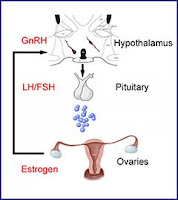
GnRH analogues for prevention of premature ovarian failure


Various glomerulonephritides unfortunately often require treatment with chemotherapeutic agents, such as cyclophosphamide. A potential adverse effect of these agents in females of child-bearing age, is premature ovarian failure. Nate previously posted about this. Since then, there have been a couple of papers published that address this dilemma further.


A meta-analysis of six randomized controlled trials reported that GnRH treated women had a higher odds of return of spontaneous menstruation than those not treated (OR 3.46; 95% CI 1.13 – 10.57), but this did not translate into a significant difference in the rate of spontaneous pregnancy after chemotherapy. However, many people are cautious in the interpretation of these results, given the small sample size in the trials, heterogeneity of protocols and varying follow-up times.


More recently, a randomized trial of triptorelin vs standard care in women with breast cancer was published. This study, performed in Italy, randomized 133 women to GnRH and 148 to standard care. Subjects were reviewed one year after the last cycle of chemotherapy for occurrence of early menopause (defined as no resumption of menstrual bleeding and postmenopausal levels of FSH and estradiol one year after cessation of chemotherapy).
The rate of early menopause was 25.9% in standard care vs 8.9% in the GnRH group (p for difference <0.001).


Whilst encouraging, these findings must be tempered by the fact that recovery of menses does not directly translate to fertility (limited data was available for this cohort at the time of publication). Furthermore, the chemotherapeutic regimens used, dosing and the duration of treatment make it hard to generalize the findings to a ‘nephrology disease’ population. There are alternative methods available, with somewhat stronger recommendations from experts in the field – these include embryonic cryopreservation.

Wednesday, April 27, 2011

Post-transplant malignancy 101

What are the most common type of cancers in kidney transplant recipients?

The three most common cancers are: non-melanoma skin cancers, Kaposi’s sarcoma and non-Hodgkin lymphomas. Compared to the general population, the risk of squamous cell carcinoma is 100x greater in transplant recipients. One interesting aspect about transplant recipients is that they have a much higher risk for a few specific malignancies but are not broadly predisposed to all cancers. As an example, the incidence of most common solid tumors in the general population (lung, prostate, breasts and colorectal) is only modestly increased or similar to the transplanted population. This suggests that other factors might play a role in the pathogenesis of malignancy post-transplantation.

What is the cause of increased risk of malignancies after transplantation?

There are several factors that seem to be associated with the development of malignancy after transplantation: impaired immune surveillance secondary to immunosuppression; carcinogenic factors like sun exposure; and host factors such as genetic predisposition to cancer, presence of oncogenic viral infection and prolonged dialysis.

How does a viral infection may increase the risk of malignancy?

Certain virus like EBV, HHV-8 and HPV carry viral oncogenes in their genome, which directly affect cell-cycle and apoptosis regulation. For example, HPV has the E6 oncogene that produces a protein that binds to p53, a major regulator of cell growth and tumor suppressor protein. Following E6 binding of p53, p53 is degraded, allowing unchecked cellular cycling and accumulation of chromosomal mutations without DNA repair.

Do different immunosuppressive drugs vary in their risk of post-transplant malignancy?

In general, the intensity and duration of immunosuppression therapy plays a major role in determining this risk of malignancy, since it severely disrupts the immune surveillance of cancers cells and dampen anti-viral immunity. Moreover, certain specific drugs like azathioprine and cyclosporine have been shown to directly lower the repair rate of DNA damage induced by solar UV radiation. In contrary, mTOR inhibitors have been shown to inhibit tumor growth mediated by both blockade of the PI3K-Akt-mTOR pathway, which is frequently activated in cancer, and by angiogenesis inhibition.

Compared to the general population, are there special screenings required for the kidney transplant recipient?

Since skin cancer is one of the most prevalent malignancies, recipients should be educated about their increased risk and recommended to reduce levels of sun exposure (especially between 10am and 4pm), wear protective clothing and apply sunscreen to sun-exposed areas, perform self-examinations and annual skin examinations by qualified health professional. For non-skin cancers, the KDIGO guidelines recommend solely reinforcement of the regular cancer screenings performed in the general population.

If a malignancy arise, what is the ideal approach?

The cancer should be managed with specific therapies for the particular tumor type associated with reduction and/or conversion to a mTOR inhibitor. The conversion to mTOR inhibitors have been successful in inducing complete regression of Kaposi’s sarcoma (figure) and decreasing the recurrence of skin cancers. General contraindications for conversion in kidney transplant recipients are proteinuria greater than 500mg/day and GFR less than 40 ml/min.

Gap in knowledge: we still need some prospective trials in order to determine if further cancer screening exams would be warranted in the transplant population that carries other competing risks like cardiovascular disease and infection.