Showing posts with label acute tubular necrosis. Show all posts

Monday, December 10, 2012

Image of the Month - Pathology

A 60 year-old woman with a longstanding history of hypertension, scleroderma and MGUS presented to the emergency room with diarrhea, vomiting and AKI (creatinine increased from baseline of 1.5mg/dl to 3.1mg/dl). She had a distant history of membranous nephropathy diagnosed on a renal biopsy 25 years previously. She had been worked up in the renal clinic for CKD and had a renal US showing relatively small kidneys. Her medications included an ACE inhibitor.

On admission, she had no hematuria. Her BP was elevated although she had a significant postural drop. She had dipstick proteinuria and she was empirically started on oral steroids for a possible GN. A renal biopsy was performed:


The image above is of the renal cortex. A single glomerulus is seen (A) which is hypoperfused. There is significant dilatation of the tubules (B) indicating acute tubular injury. There is attenuation and degeneration of the tubular epithelial cell layer. At the lower end of the image is an atrophic tubule with a hyaline cast.


This image again shows a hypoperfused glomerulus. However, the main finding here is an extremely damaged arteriole (A). There is multilayering within wall of the vessel (that was replicated throughout the biopsy). The vascular lumen is almost occluded with endocapillary proliferation and remodeling of the vessel wall.

In some areas of the biopsy (better perfused), the glomeruli looked relatively normal. There was some mesangial expansion with irregular capillary loops but no evidence of membranous disease.


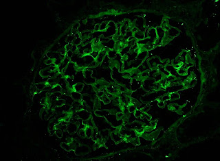
IF showed some minimal deposition of IgG in the mesangium but equal kappa and lamda light chains.


Finally, the EM showed multiple mesangial deposits (A) of uncertain significance but again, no evidence of membranous disease. Notably, there were was no evidence of active inflammation in the biopsy.

Following the report of the biopsy, the steroids were stopped. The patient's blood pressure was controlled and her renal function returned to baseline within a few days. It is likely that this acute episode was related to volume depletion and acute tubular injury exacerbated by her severe underlying vascular disease. Interestingly, she now has no albuminuria and she is back on her ACEi. The significance of the mesangial deposits remains unclear and she will be followed on an ongoing basis in the renal clinic.

We often see patients like this on consult who present with AKI following a GI illness while on an ACEi. However, we don't normally get to see the pathology in these very common case.

Click on any image to enlarge

Tuesday, August 23, 2011

AKI requiring dialysis in the hospital: Does this mean I'm on dialysis for good?

As nephrology fellows we see a lot of Acute Kidney Injury (AKI) on the inpatient consultative service. When AKI in the hospital requires dialysis people and their families naturally want to know whether this means long term dialysis will be required.


The answer, as with many things in medicine, is it depends. AKI has a wide variety of causes but one of the most commonly encountered entities in the hospital is Acute Tubular Necrosis (ATN) and luckily we have a number of good studies to help inform prognosis.


One of the first things to realize about hospitalized AKI due to ATN that requires dialysis is that it is associated with a very high in-hospital mortality rate. In the ATN study which examined critically ill ICU patients with AKI presumed secondary to ATN randomized to either intensive or less-intensive dialysis there was a roughly 50% in-hospital mortality in both groups.


In those with ATN who require dialysis and survive hospitalization, whether or not long term dialysis is required depends on the demographic. In general, the lower the baseline kidney function at the time of AKI the higher the rates of long term dialysis dependence.


The ATN study excluded patient's with advanced CKD (about 30% of patients though had moderate CKD with GFRs between 30 and 59 ml/min/1.73m2). Around 70% of people who survived to day 28 continued to require dialysis. In contrast, in a German cohort of 433 critically ill patients all with GFRs of greater than 90 ml/min/1.73m2 who developed dialysis requiring AKI from ATN not one survivor (in hospital mortality was again approximately 50%) required long term dialysis at discharge.


In terms of more advanced CKD, a study using the Northern California Kaiser Database looked at patients both in and out of the ICU who developed AKI requiring dialysis (greater than 90% of patient's had likely ATN by subset chart review). Of those that survived the hospitalization (overall mortality was 26%) 42%, 63% and 90% of patients with eGFRs of 30-44, 15-29 and less than 15 ml/min/1.73m2 respectively were felt to be dialysis dependent within 30 days of hospital discharge (a selected chart review revealed no cases of recovery within three months).


In summary those with AKI secondary to ATN who require dialysis in the ICU have a very high mortality rate. Of survivors, approximately 70% will require long term dialysis unless they enter the hospital with completely normal renal function in which case the chances of renal recovery appear to be quite good. Patient's with the most advanced form of CKD who develop AKI requiring dialysis are very unlikely to recover.

Saturday, December 19, 2009

Ischemia-Reperfusion Models

The study of AKI/ATN has relied heavily on one particular animal model: the warm ischemia-reflow model (often referred to as "ischemia-reperfusion injury"), in which one of the renal arteries is transiently ligated off for a set period of time while body temperature is maintained, then opened up and allowed to reperfuse the kidney. A recent review in Kidney International by Heyman et al addresses some of the limitations of this model, mostly in terms of differences between the mouse model of ischemia-reperfusion and the typical human AKI/ATN we experience clinically.

Some the important differences between mouse and human AKI: First, while the warm ischemia-reperfusion model tends to initially target the S3 segment of the proximal tubule and typically leads to overt tubular necrosis, in human AKI necrosis is not always present, and when it is tends to be patchy and most commonly affecting the distal nephron (in particular: the medullary thick ascending limb and medullary collecting ducts). Second, in clinical practice it is COLD ischemia which is often the mechanism of injury (e.g., surgical procedures in which the aorta is cross-clamped is often performed in the setting of a lowered core temperature, and donated kidneys are typically stored on ice prior to transplantation), rather than warm ischemia. Overall, the authors conceded that the rodent ischemia-reperfusion model has been invaluable, but caution against using it to explain all aspects of human AKI/ATN.

The notion of whether AKI/ATN occurs primarily in the proximal versus the distal tubule is not merely of academic interest. According to one version of the "distal nephron model", the reduced GFR experienced in response to medullary hypoxia is actually an adaptive response: decreasing the metabolic demands of tubular epithelia would decrease hypoxic injury; in a sense one could think of the nephrons as "hibernating" in a low metabolic state until they sense that the hypoxic insult has been removed and they can resume optimal cellular function. If this is true, it might caution physicians from trying to stimulate GFR in patients with AKI, instead encouraging attempts to limit tubular energy expenditure.

Saturday, November 14, 2009

Aminoglycoside Toxicity

The aminoglycosides are amongst the most well-known nephrotoxic drugs. Yet due to their efficacy against many organisms, they are amongst the most common antibiotics used. The next time your nephrology service goes toe-to-toe with the infectious disease service in the ongoing battle as to whether or not a patient with CKD should get gentamicin, you can arm yourself with these factoids regarding the mechanism of aminoglycoside toxicity:

Aminoglycosides are a potent tubular toxin; the reduction in GFR which results is therefore thought to be an indirect effect on the glomerulus. The predominant sites of aminoglycoside toxicity are the S1 & S2 segments of the proximal tubule. Aminoglycosides are filtered by the glomerulus, and once concentrated in the urine in these segments occurs, they are known to bind to phospholipids, followed by internalization within the cell via megalin. Once inside the proximal tubular cell, they they are concentrated within lysosomes and cause a stereotypical disorganization of the lysosomes termed "myeloid bodies."

The myeloid bodies are a sign that the tubular cells are functioning poorly, and as a result there is decreased tubular function often manifesting as K, Mg, Ca, PO4, and glucose wasting--almost like a "Fanconi's Syndrome" picture. Overt necrosis of the tubular epithelial cells can also occur, resulting in ATN. Stereotypically, aminoglycoside toxicity results in non-oliguric renal failure which more often than not recovers (after a few weeks) once the drug is withdrawn.

From a pharmacokinetics standpoint, the toxicity of aminoglycosides correlates best with the peak concentration of the drug. Interestingly, in common clinical practice the drug is dosed based on following aminoglycoside trough levels...

Tuesday, March 3, 2009

There's No "N" in ATN

Just like there's no "I" in team...there's no "N" in ATN.  

I heard this expression at our Renal Grand Rounds today featuring pathologist Isaac Stillman at nearby Beth Israel Hospital.  It's not completely accurate, as in many cases of ATN, one can often see patchy loss of tubular epithelial cells with resultant gaps and exposure of denuded basement membrane--though recent evidence points to the fact that this is can be the result of either necrosis or apoptosis, a more controlled version of cell death.  

But it is true that, despite its name, frank necrosis of renal tubular cells on biopsy is actually fairly rare.  ATN remains predominantly a clinical diagnosis, and there are several instances in the literature of patients with a clear diagnosis of ATN who, on renal biopsy, demonstrate a seemingly normal renal histology.  Most likely, there is tubular injury, but we just don't have the sensitivity to detect it.  This is supported by recent animal studies identifying various renal biomarkers which are elevated in the urine of virtually all mice subjected to AKI despite apparently normal tubular histology.  

When there is necrosis in ATN, it is often most prominent in the outer medulla, where the S3 segment of the proximal tubule joins the medullary thick ascending limb of the loop of Henle, as this is a region already subjected to relative hypoxia.  Heavy metal-induced ATN is a special subset of ATN known to demonstrate especially high levels of necrosis than the more common forms of ATN we typically see.    

Wednesday, January 21, 2009

Do Kidney Stem Cells Exist?

Hot topic of research: do kidney stem cells kidney exist?

Stem cells are defined as a subpopulation of cells which retain the ability of self-renewal and differentiation into a specialized cell type. Stem cells specific to many tissue types have been identified (e.g., hematopoietic stem cells, brain stem cells, etc) and the question arises: are there kidney stem cells?

Obviously, the renal tubular epithelium is capable of regeneration: the majority of patients who experience ATN will regain renal function as a result of a robust regenerative response which repopulates the denuded basement membrane with new tubular epithelial cells over time. There are three possible sources of new tubular cells:

1. Adjacent, less damaged tubular cells could repopulate the epithelium under the right conditions.

2. Circulating, bone marrow-derived stem cells may exist.

3. "Resident kidney stem cells" refers to the possibility of a subpopulation niche of renal cells that lives in the kidney which could be responsible for tubular regeneration when required.

Thus far, the evidence seems to point to (1)--that all renal epithelial tubule cells, if they can survive the injury, have the capacity to repopulate the tubular epithelium. Perhaps understanding this pathway more completely would allow us to generate new pharmacologic therapies to stimulate or hasten the repair response.

It is important also to point out that this field is still young, and perhaps there really are "kidney stem cells" that we don't have the technology to detect yet. Resident kidney stem cells do appear to exist in other organisms, such as some fish.

Tuesday, December 30, 2008

Biomarkers: The Race for a Better Creatinine

The Panel 7: I remember it was one of the first things I learned in clinical medicine. Na, K, Cl, HCO3, BUN, creatinine and glucose. I know not all countries use the same "grid format" of presenting these numbers as we do in the U.S., but all of them use creatinine as the indicator of GFR which is so useful in interpreting a patient's electrolyte values.

While certainly useful, there is one big problem with creatinine that should be fairly obvious to all renal fellows: it is a relatively late marker of renal injury. By the time the creatinine has risen, there has already been significant renal damage. For some surgical patients in the ICU, immediately post-operatively after a CABG for example, we will start renal replacement therapy even with relatively "normal" creatinines in the 1.3 - 1.4 mg/dL range--if we feel there is a genuine acute kidney injury, as manifested (for instance) by anuria and volume overload. What if there was a blood test which indicated renal damage at the earliest stages of AKI--e.g., a substance whose concentration in the bloodstream (or urine) became significantly elevated before that of creatinine?

This is what the field of biomarkers is all about, and the race is on. Imagine a day where instead of creatinine in the lower right hand corner of the panel 7 there is a different test. An excellent review of the field can be found here, look for the April 2008 edition of the Brigham & Women's "Nephrology Rounds" publication by Won Han. Four of the big biomarkers under investigation currently include neutrophil gelatinase associated lipocalin (NGAL), kidney injury molecule-1 (KIM-1), interleukin-18 (IL-18), and cystatin C. Many of them are detected in the urine--with the rationale that the earliest indication of tubular kidney damage would be to look for the presence of proteins expressed by renal tubular epithelial cells in response to ischemic damage in the urine. It could very well be that there is no single "winner" of the best test to detect early renal damage--perhaps different biomarkers can be used to detect injury within discrete regions of the nephron (e.g.,proximal tubular damage versus distal tubular damage), or perhaps different biomarkers will be able to detect different types of nephrotoxin-induced damage (e.g., contrast nephropathy versus sepsis-associated AKI). This is an interesting field with a promising future.

Monday, December 15, 2008

'Shrooms and Renal Failure

While toxic mushroom ingestions are generally known for their ability to cause acute liver failure, certain varieties of mushrooms can also result in acute renal failure.

One such type of mushroom is Amanita smithiana, (shown at left) which is responsible for causing relatively rapid (e.g. within a few days of ingestion) renal failure. The mechanism is felt to be ATN and there have been several case reports of this type of mushroom ingestion in the Pacific Northwest region of the United States.

Another distinct type of mushroom-induced acute renal failure is those produced by Cortinarius species (shown at bottom). These are found mostly in Europe, and the mechanism of renal injury here is a tubulointerstitial nephritis. As such, onset can be delayed (3 days - 3 weeks) and often less severe than Amanita-induced renal toxicity.

Also, as liver failure is still the most common disease associated with toxic mushroom ingestion, hepatorenal syndrome (as a secondary event) must also need to be strongly considered in an individual with renal failure following mushroom ingestion.


Friday, December 5, 2008

cisplatin-induced acute kidney injury

At the Brigham and Women's Hospital & the Dana Farber Cancer Institute, there is an ongoing trial of using pneumonectomy and injected, heated cisplatin into the pleural space for patients with malignant mesothelioma, a condition with an overall poor oncologic prognosis and limited treatment options.  Apparently this technique has had some success, but not surprisingly, it results in many renal consults to the Nephrology Fellow as a very substantial percentage of patients have significant acute kidney injury and many require dialysis.  Unlike standard chemotherapy where cisplatin may be held once renal toxicity has occurred, the injected cisplatin has a very slow, continuous absorption and therefore there is no opportunity for reversal once this sets in.  

The mechanisms of cisplatin-induced acute kidney injury are still being worked out but generally result in tubular toxicity (ATN).  Both necrosis as well as apoptosis appear to be involved, according to this recent KI review article on the topic.  

Cisplatin falls under the category of platinum-containing alkylating agents.  Related drugs include carboplatin and oxaloplatin, which still have some renal toxicity but substantially less than cisplatin.  

Sunday, November 30, 2008

Scoring System for ATN vrs pre-renal AKI

One of the earliest time-saving tricks I learned as a Nephrology fellow was to always ask the covering housestaff to have a specimen of piping hot urine waiting for me at the time of the initial consult:  I was trained to do a urinalysis on all patients on whom I am called to see.  While I have been impressed with its usefulness of several occasions, there is surprisingly little standardization for the technique, and not a lot of literature providing objective criteria for diagnosis.

A recent study in C-JASN by Perazella et al looked at the ability of the urinalysis to distinguish between ATN versus pre-renal AKI on 267 consecutive inpatients with AKI in the Yale-New Haven Hospital system on whom a renal consult was called.  Participating nephrologists were asked to assess a cause of either "ATN", "pre-renal", or "other" at two different time points:  (1) just after history & physical but before microscopy diagnosis, and (2) after patient discharge or death.  As biopsies were not done in this study, the 2nd & final diagnosis--based on clinical judgement--was the "gold standard."  The urinalysis itself was carried out in a standardized fashion and a scoring system based on the number of casts or renal tubular epithelial cells (RTECs) was developed.

Not surprisingly, patients with a high "urine sediment score" (indicating many casts or RTECs) had a high rate of ATN as a final diagnosis while those with lower scores were more likely to have pre-renal AKI as a final diagnosis.  While this is not groundbreaking, what is worthwhile about this study is that (a) they prove that the urinalysis is a valuable tool for diagnosis by showing that a significant number of patients had their diagnosis changed following urinalysis, and (b) they tackle this problem in a systematic fashion.

Tuesday, May 20, 2008

ATN Study: More Is Not Better

The results from the Acute Renal Failure Trial Network ("ATN Study") are revealed in the most recent issue of the New England Journal of Medicine.

The study was a randomized control trial in which ICU patients with acute kidney injury were randomly selected to receive either standard-dosed dialysis (defined as three times a week hemodialysis or CVVH at 20 cc/kg/hr) or more intensively-dosed dialysis (defined as six times a week hemodialysis or CVVH at 35 cc/kg/hr). The trial was not, as some people erroneously believe, intended to settle the contentious issues of whether there is any benefit of CVVH over intermittent hemodialysis; patients were actually permitted to move back and forth between intermittent hemodialysis and CVVH provided they stayed within the intensive versus the standard group to which they were originally assigned.
.
The results: no significant difference was found in the standard compared to the intensively-dosed groups. The study is somewhat at odds to the famous Ronco study published in 2000 in the Lancet which demonstrated a survival benefit in patients who received a higher dose of CVVH (either 35 or 45 cc/kg/hr) compared to those receiving a lower dose (25cc/kg/hr).