Showing posts with label Paul Phelan. Show all posts

Friday, March 11, 2016

#NephMadness 16: Let’s win it for the Fellows


NephMadness 2016

It’s finally arrived, the fourth annual NephMadness has dropped with 32 new Nephrology concepts battling it out for the month of March to see who will be crowned champion. Fully referenced blog posts on each of the topics are available at AJKD Blog and an explanation of the event can be found here for our new participants. Essentially, it involves reading up on the content provided and deciding which Nephrology concepts win in head to head, knockout match-play. You may fill out your bracket (pick your winners) using our online tournament page until March 23rd.

I’m hoping this year the winning participant will be a fellow/resident (or SpR/SHO) or better still a group of trainees. Last year there was a trend towards fellowship programs coming together, debating the topics and matchups and making their picks as a team. This is a fantastic way to extend the free online medical content to real-world flipped classroom teaching sessions at your institution. I’d also love to see this extend outside of the US to SpR training programs in Europe, Asia and beyond. So I propose you enter the contest individually but also with your colleagues as a group. Eternal glory awaits!

Thursday, February 26, 2015

#NephMadness 2015: It’s almost here

It’s almost March, time to fill out your bracket. No not college hoops, it’s time for the annual Nephrology SoMed educational phenomenon that is NephMadness. The brainchild of Matt Sparks and Joel Topf has grown legs in its 3 years of existence and now represents a highlight of the Nephrology #FOAM calendar.

NephMadness is a homage to the NCAA Basketball Tournament, March Madness, but instead of matching up college basketball teams, NephMadness throws some of the most important concepts in nephrology together to battle it out. This years theme is Nephrologys interaction and cross-over with other specialties. See the current editorial in AJKD by the NephMadness team for the complete low-down. The overall regions/specialties, each with 8 Nephrology topics, for 2015 are:

1.         Obstetric Nephrology
2.         Infectious Disease and Nephrology
3.         The Heart and Kidney Connection
4.         Nephrology and Nutrition
5.         Genetic Nephrology
6.         Critical Care Nephrology
7.         Nephrology and Vascular Surgery
8.         Onconephrology

The game will progress throughout March with winners and losers announced along the way via ongoing blog posts. My own (extremely biased) view is that the winner will come from the strong Genetic Nephrology region! Let us know what you think when the brackets are published on March 1 on the AKJD blog. Also follow along on Twitter using the hashtag #NephMadness.


Thursday, November 6, 2014

NephJC Live at ASN Kidney Week 2014

The Nephrology twitter journal club will come to life on Saturday 15th November at Kidney Week in Philadelphia. NephJC co-founders Dr Joel Topf and Dr Swapnil Hiremath will co-host the event at the Double Tree hotel.

There will be 2 live presentations on the day, both of which are sure to stimulate active discussion. There will be a study, presented by Dr. Deirdre Sawinski of University of Pennsylvania, looking at transplanting HIV positive kidneys into seropositive donors. Dr Francis Wilson will also present his RCT on acute kidney injury.

The event is filling up so please visit www.nephjc.com before this Sunday November 9th to register. For those of us who cannot make it to Philadelphia, the event will, as ever, be live tweeted using #NephJC.

Thursday, August 21, 2014

Renal Function after Off- or On-Pump CABG: CORONARY Trial is next #NephJC

The next Nephrology online journal club (#NephJC) will discuss the results of the CORONARY Trial, presented at the late breaking session at the ASN and published this year in JAMA. The trial compared patients undergoing their first coronary artery bypass graft (CABG) surgery using an off- or on-pump technique. The main study published previously revealed no difference with respect to the composite outcome of 30-day mortality, myocardial infarction, stroke or acute kidney injury (AKI) requiring dialysis. The renal function trial was a prespecified substudy involving 2975 (of a total 4752) consecutive patients enrolled in CORONARY with baseline and post-operative serum creatinine data. The renal substudy patients had similar characteristics to the overall CORONARY population.

Outcomes of Interest:
  • Post-operative AKI was defined as a 50% increase in the serum creatinine concentration within 30 days of surgery (highest creatinine within 30 days was used).
  • Loss of renal function at 1 year = 20% loss in eGFR (using CKD-EPI).
Patients:
  • Worldwide enrolment with 42% from Asia and the remainder mostly from Europe (21%) and the Americas (<1% were African American).
  • Baseline characteristics of note were a mean age of 68 years, BMI 27, >80% male, almost half were diabetic and a similar number of ‘urgent’ cases between the groups.
  • Almost a quarter had CKD (eGFR <60mls/min) and the mean eGFR was 74-75mls/min in the 2 groups.
Results:
  • There were 561 AKI events (median time of 2 days post-op to peak creatinine) with a reduced rate with off-pump (17.5%) V. on-pump (20.8%) surgery (adjusted RR 0.83 [CI 0.72-0.97]; p = 0.01).
  • Mean eGFR at 1 year was 72 mL/min with off-pump and 73 mL/min with on-pump.
  • No significant difference in loss of eGFR at 1 year between off-pump (17.1%) V. on-pump (15.3%) surgery (P = 0.23).
  • Those with CKD derived a greater benefit in reduced AKI with off-pump surgery but eGFR loss at 1 year remained insignificant.
  • Over 200 patients crossed over between the groups (evenly split) and results of the intention to treat were similar to as-treated analysis.
  • Multiple alternative definitions of AKI & loss of kidney function did not alter the main results.
Discussion:
Dialysis requiring AKI has detrimental effects on long-term kidney function. Less severe AKI is more common with major cardiac surgery (only just >1% had AKI requiring dialysis in the original CORONARY trial). It is less clear what effect these more subtle derangements have on long-term function. This study suggests that these ‘mild’ AKI events may not have much longer-term significance, contrary to observational studies [ref, ref]. As pointed out by the authors, this finding has implications for other interventions in mild AKI such as for contrast nephropathy. Does preventing a subtle GFR dip in this scenario have a long-term benefit? The study is limited somewhat by the unique situation studied in the trial (although cardiac surgery provides a very ‘convenient’ insult in which to study AKI). Also, we are relying on serum creatinine and all its limitation to assess kidney function. Moreover, not all eligible patients had creatinine values measured and single measurements and imputed values were often used for the analysis.

Verdict:
This study provides good evidence that off-pump CABG decreases the rate of non-severe AKI but that this does not appear to translate into better renal function at 1 year. When I first heard the results of this study at the ASN (a somewhat deflating session along with lots of other negative/inconclusive Nephrology studies), I was disappointed with the small magnitude of the AKI decrease with off-pump surgery. I had expected the toxic milieu associated with on-pump surgery (aortic cross clamping, exposure to bypass circuit, changes in blood pulsatility) to be associated with much higher rates of AKI, compared to off-pump. The study also questions my preheld assumption that acute drops in GFR, from mild to severe, had a continuous magnitude of impact on long term renal function.
Feel free to get involved by joining the live Twitter chat on Tuesday 26th August at 9pm Eastern using #NephJC. Also, check out www.nephjc.com for more background and past journal clubs.

Tuesday, July 15, 2014

Lithium and the Kidney: Old Observations & New Insights

Lithium is indispensable as an effective treatment for bipolar affective disorder. However, it has a narrow therapeutic index with desired therapeutic levels between 0.6 and 1.2 mEq/L. Lithium is handled by the kidney (responsible for almost all its excretion) in a manner very similar to sodium. It has a molecular weight of 7 daltons, has a moderate volume of distribution, is <10% protein bound and is therefore readily dialyzable. Lithium may cause of myriad of renal related toxicities, some of which are classic descriptions and some which are less well known. Much of this has been discussed on RFN before (here, here) but I felt a refresher with some additions was due.

Nephrogenic Diabetes Insipidus (NDI)

This is perhaps the best know and most common complication of lithium therapy, with an estimated prevalence of 20-70%. Patients present with polyuria and polydipsia due to a urinary concentrating defect that can lead to volume depletion, especially if access to free water is restricted. Lithium downregulates aquaporin-2, vasopressin-activated water channels expressed on the collecting duct principal cells. It is worth noting that ongoing volume depletion from NDI may aggravate the risk of supra-therapeutic levels and therefore chronic nephrotoxicity. Amiloride may help as it inhibits lithium reabsorption via ENaC in the collecting duct.    

Renal tubular Acidosis (RTA)

Lithium may induce a hyperchloremic non-anion gap metabolic acidosis, analogous to a distal RTA. It is likely due to decreased distal proton secretion. Unlike acidosis associated with amiloride, it is not associated with hyperkalemia probably because of their different effects on distal H+/K+-ATPase function.

Acute Lithium Nephrotoxicity

This may occur as an overdose in newly treated patients or those on long-term lithium therapy. Acute lithium intoxication may cause altered mental status and acute kidney injury. Drugs which decrease GFR (NSAIDs/RAAS inhibitors) may exacerbate intoxication as can thiazides, which cause a natriuresis and a subsequent reabsorption of Na (and lithium). With preserved renal function and mild intoxication, increasing urine output/forced diuresis may be all that is needed. Care should be taken using 0.9%NaCl if hypernatremia from NDI is present. As mentioned above, lithium is readily dialyzable and hemodialysis (ideally using a high flux membrane to aid clearance) is the primary management for severe cases. A lithium level >4mEq/L is considered an absolute indication for dialysis in most cases. A level >2.5mEq/L with severe symptoms, or renal impairment which will slow native clearance, is also an indication. If in doubt, most nephrologists would have a low threshold for dialysis, although it is usually not needed with a level <2.5mEq/L. Drug levels drop rapidly during dialysis but they do rebound so extended and repeated sessions are usually necessary. Continuous therapies are less efficient so are not desirable if severe intoxication is present but may be used if conventional dialysis is not immediately available.

Chronic Tubulointerstitial Nephritis (TIN)

A chronic TIN with interstitial fibrosis and tubular dropout is the commonest pathology seen when patients with lithium and renal impairment are biopsied. Consistent with this is a bland urine sediment with little/no proteinuria being common (although not exclusive-see next section). As nephrologists, our view of the prevalence of lithium induced CKD is skewed. The exact prevalence is difficult to determine but many patients have preserved renal function. The tricky decision is whether (and if so when) to stop therapy? This is rarely a decision the nephrologist can make alone and should be balanced with efficacy of treatment/alternatives available and usually led by the psychiatrist in my opinion.

Nephrotic Syndrome

It is less well known that lithium may be associated with a glomerulopathy. Minimal change disease is most often described but membranous nephropathy and FSGS has also been reported. Cases were considered lithium induced as proteinuria disappeared upon cessation of the drug, and when re-challenged (for psychiatric reasons), the nephrotic syndrome recurred. A series of 24 patients with lithium-induced nephrotoxicity from New York included one quarter with nephrotic-range proteinuria. My initial thoughts were that the FSGS lesions were likely secondary to nephron loss and resulting hyperfiltration injury. However, arguing against this is that presence of FSGS lesions did not correlate with severity of tubulointerstitial lesions. Moreover, there was a high incidence of extensive podocyte foot process effacement, to a degree uncommon in secondary FSGS. The mechanism of glomerular toxicity is unclear.

Renal Cell Tumors

It has recently been reported in Kidney International that chronic lithium use is associated with an increased risk of kidney tumours.  After a mean duration of lithium exposure of >21 years, patients had a significantly higher risk of solid renal tumours compared with gender, age and eGFR matched controls [Standardized Incidence Ratio i.e. ratio of observed-to-expected numbers of renal cancers was 7.51 and 13.69 in men and women respectively]. The tumours were a mixed bag of benign (oncocytomas, angiomyolipomas etc.) and malignant (clear cell, stromal, papillary) lesions. While these results are thought provoking, this was a retrospective study and the design seems open to detection bias in my opinion (were patients on lithium screened more than regular individuals?).

Hyperparathyroidism

As previously posted, lithium may cause hypercalcemia and stimulate PTH via a variety of postulated mechanisms (see previous post).


Friday, July 11, 2014

Parvovirus B19 and the Kidney

Parvovirus B19 (PVB19) is a small, non-enveloped, single-stranded DNA virus belonging to the Erythrovirus genus, named for their tropism for erythroid precursor cells. It is the only known parvovirus to infect humans. By late adulthood, most people have IgG Anti-PVB19 serology demonstrating previous exposure, often from an asymptomatic infection. However, infection with PVB19 may cause a variety of clinical syndromes including fifth disease (‘Erythema infectiosum‘, a childhood viral exanthem), a polyarthropathy, pure red cell aplasia and hydrops fetalis in utero. The nephrologist may encounter PVB19 in 3 broad settings: 

Post-Transplant 

The incidence of PVB19 infection post kidney transplantation is hard to accurately determine but is likely in the range of 1-10%. Infection tends to occur in the first year and frequently in the first few weeks, suggesting possible donor transmission but the mechanism of infection/trans mission is unknown. Clinical syndromes are mainly acute anemia and chronic pure red cell aplasia, although a pancytopenia may develop. The few cases I have seen have been easy to identify, as patient presented with a profound isolated anemia. The diagnosis of chronic anemia or pancytopenia may be more protracted as these are obviously common post-transplant complications (graft dysfunction; drugs-immunosuppressants, anti-virals, co-trimoxazole, ACE inhibitor; other infections-CMV etc.). Treatment consists of reducing immunosuppression (usually the anti-metabolite) and IVIg. Similar to other viral infections (CMV, Polyoma viruses), there is some thought that PVB19 may be associated with allograft dysfunction or even acute rejection (?injured endothelium exposing previously hidden epitopes; no hard evidence for this however). A case series of thrombotic microangiopathy with PVB19 infection post transplant isolated PVB19 DNA from graft biopsies, however, overall the evidence for PVB19-induced allograft dysfunction is weak at present. 

Glomerular Disease 

PVB19 may cause a variety of glomerular lesions in immunocompetent hosts. These were first noted as associations with PVB19 viremia, however, subsequently viral DNA has been identified in renal cells from biopsy tissue. The most well described pattern of injury is FSGS, particular a collapsing glomerulopathy (see image), where the PVB19 genome has been detected in podocytes and parietal epithelial cells by PCR. Proliferative glomerulonephritis has also been temporally associated with PVB19 infection. A syndrome similar to acute post-infectious glomerulonephritis may occur, displaying hypocomplementemia, endocapillary and mesangial proliferation with subendothelial electron dense deposits. Acute glomerulonephritis may be more common in patients with sickle cell disease after an aplastic crisis due to PVB19. Given the ubiquitous nature of PVB19, it is likely that host factors contribute to why certain individuals manifest glomerular disease, with genetic factors, as always, being implicated. Viral DNA may be found in renal tissue after the acute infection has passed and the glomerular lesions will not necessarily improve with resolution of the infection. 

Dialysis Patients 

Patient with ESRD are considered to be at risk for aplastic crisis due to PVB19, albeit with a paucity of data to support that claim. The presence of abundant immature erythroid cells (due to EPO use), a relative immunosuppressed state and some particular patient populations (e.g. sickle cell disease) lead to an increased theoretical risk of this complication. 

PVB19 may be diagnosed using serology although immunosuppressed patients may not mount an adequate antibody response so PCR viral load is commonly employed. Treatment is non-specific and is generally supportive. IVIg is often used as these pooled preparations have anti-PVB19 antibodies, although randomized controlled data supporting its use is not available. Cutting back immunosuppression in transplant patients may also be of benefit, initially by reducing/stopping the anti-metabolite (logical as mycophenolate frequently induces bone marrow suppression independently). Also, tacrolimus is considered by some to be particularly conducive to PVB19 infection and a switch to cyclosporine may be a next step. 
To summarize, PVB19 is important to the nephrologist from a clinical perspective particularly for its tendency to cause isolated severe anemia (or pancytopenia) in our immunosuppressed patients. It may also cause glomerular disease in immunocompetent patients and should be a differential diagnosis for otherwise unexplained glomerulopathy, especially collapsing FSGS. It also provides clues from a research perspective to mechanism of glomerular disease/sclerosis.

Monday, June 23, 2014

IgM Nephropathy: What You Need to Know


Similar to my recent post on C1q Nephropathy, IgM Nephropathy (IgMN) is a podocytopathy that is often considered a variant of Minimal Change disease (MCD) or FSGS. Like C1QN, it is also a controversial entity with a lack of uniformity from a diagnostic point of view. This undoubtedly leads to under-reporting with some pathologists calling a case IgMN what others would call MCD/FSGS/Mesangial Proflierative GN with low level IgM staining.
First descriptions of IgMN reported mesangial hypercellularity on light microscopy, granular IgM and C3 mesangial deposits on immunofluorescence with electron dense deposits in about half of cases. Podocyte foot process effacement was also evident consistent with the presentation of nephrotic syndrome. The incidence has been reported to be 2-18% of renal biopsy series and age at presentation appears to be bimodal with a peak in childhood and again later in life, 6-7th decades. The incidence of IgMN may be higher in the developing world and most recent reports come from developing nations (ref, ref).
The accurate diagnosis of IgMN is important from a prognostic and therapeutic point of view. Overall, IgMN does not respond to steroids as well as MCD with a higher proportion of patient with steroid-dependent and resistant disease. Furthermore, assessment of children with steroid-dependent and resistant MCD revealed a high proportion of IgMN, which appeared to respond better to cyclosporin than to cyclophosphamide. Rituximab (as ever!) may be another alternative therapeutic choice, although evidence remains at case report level, including a case of recurrent IgMN post-transplant. Similar to C1QN, the severity is likely dependent on the light microscopic changes with MCD patterns reporting an excellent remission rate and an FSGS pattern of injury having a worse prognosis (ref). Most cases however may have mesangial proliferative changes evident. There is even a report of IgMN presenting with crescentic lesions.

IgM deposition may be seen in many other glomerular disorders and its role is unclear. Is it an innocent bystander, reflecting natural IgM binding to exposed epitopes on injured glomerular cells? There is some evidence that IgM deposition may activate complement and that efforts to prevent IgM antibody deposition may prevent complement activation and slow the progression of glomerular injury. IgMN is another glomerular disease that we have an unclear understanding of pathogenesis and etiology where we rely on crude pathological descriptions. Hopefully the future will bring clarity to these diseases and facilitate precise molecular diagnosis of entities such as IgMN.

Sunday, June 22, 2014

POSEIDON Trial for Contrast Induced Nephropathy: The Next #NephJC

The next Nephrology Twitter Journal Club (@NephJC) will discuss the POSEIDON Trial which has been published in The Lancet. Interest in the prevention of Contrast Induced Nephropathy (CIN) is alive and well with several novel approaches/therapies being recently reported (see my recent post), mostly in non-Nephrology journals. Despite these interventions, volume expansion remains the cornerstone of preventative management for CIN (contrast-induced acute kidney injury is just too much of a mouthful for me).

This current trial randomized patients into 2 protocols of 0.9% NaCl-based volume expansion peri-cardiac catheterization. The study group had their fluid rate governed by left ventricular end diastolic volume, measured easily during the catheterization procedure and used as a surrogate for volume status. This allowed the intervention group patients to safely receive higher volumes of saline during their procedures which translated into significantly lower rates of CIN. Obviously this method can only be conveniently employed during catheterization procedures and would need the involvement of interested cardiologists for it to be used in real-life clinical practice. However, the results appear impressive.
You can read a concise summary of the trial and results over at NephJC. Be sure to tune into Twitter on Wednesday June 25th at 9pm Eastern and feel free to get involved using #NephJC.

Saturday, June 14, 2014

C1q Nephropathy: What You Need to Know

I saw a 20 year old African American male in clinic with nephrotic range proteinuria, reduced GFR and no signs or symptoms of a systemic disease. His mother developed ESRD at age 36 and his brother, aged 26, has CKD Stage 4. A full laboratory workup including complement proteins and ANA was unremarkable. Renal biopsy demonstrated an FSGS pattern of injury with a full house picture on immunofluorescence but with dominant C1q staining. A diagnosis of C1q Nephropathy (C1QN) was made. 

Jennette et al described C1QN in 1985 in 15 patients without evidence of lupus who had dominant mesangial C1q deposition, often with C3 and Ig’s also. The mean age at presentation was almost 18 years, with a similar male: female ratio and average proteinuria of 7.5g/day. Another series from Columbia University included 19 patients who were predominantly African American (73%), young and female (73%) with nephrotic range proteinuria. Renal outcomes across the series’ (including this European series) are mixed but would suggest immunosuppressive treatment directed against the primary pathology should be considered.
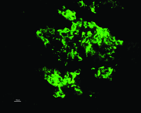
C1QN may be considered an independent pathological entity, although this has been controversial, with some classing it as a subtype of FSGS or minimal change disease (MCD). The prevalence varies (0.2-16%) but is more common in children. The predominant light microscopic appearance may be MCD, FSGS or a proliferative glomerulonephritis. While C1q deposition (predominantly in the mesangium; see image) is dominant or co-dominant by definition, a full house pattern of IgM, IgG, IgA, C1q and C3 may be observed, more commonly with a proliferative glomerulonephritis. As for treatment, the case series are small and largely uninformative. Some patients appear to remit with or without immunosuppressive treatment whereas others have a rapid course towards ESRD. Patients with an FSGS pattern may do worse than other types. For what it’s worth, my patient had no response to 6 months of steroids and mycophenolate and a plan for cyclosporine was made. The fact he likely has a genetic element to his disease adds complexity. Some would argue that immunosuppressive therapy in these cases is not likely to be of benefit but we see from our hereditary FSGS cohort that this is not necessarily the case. Cyclosporine and Rituximab in particular appear to have a directly beneficial effect on podocyte function (also see case report of Rituximab treatment in C1QN).
As for the pathogenesis and significance of the C1q deposits, we have much to learn. Are the C1q deposits pathogenic or merely markers of injury? We do know that C1q binds to the basement membrane via laminin and C1q receptors enhance binding of immune complexes to mesangial cells where it combines with other complement proteins to form C1 protease. This in theory allows activation of the classical complement cascade. Despite this, serum C4 levels are generally normal and I can find no reports of complement directed therapy (Eculizumab or C1-Inhibitors) in C1QN.
Bottom line: C1QN is likely an under recognized entity which can have varying clinical and histological presentations. Renal outcomes are also variable and it may have a different prognosis compared to traditional MCD (& FSGS?). One to keep in mind for the boards.

Sunday, May 11, 2014

The use of IVIg in Kidney Disease

It’s time for a quick nephro-centric summary of immune globulin use. Immune globulin, usually administered intravenously (IVIg), is made from pooled human plasma and used for a wide variety of human disease. It contains mostly IgG with various IgA concentrations depending on the preparation and different stabilizers (see sucrose nephropathy below). IVIg has various anti-infections and anti-inflammatory effects via mechanisms that are still incompletely understood. The sphere of renal transplantation is where most nephrologists will see it being administered.

HLA Desensitization
IVIg is incorporated into various desensitization protocols which may decrease preformed anti-HLA antibodies and render a previous positive crossmatch negative. Two broad regimes are (a) high dose IVIg at 2g/kg single dose or monthly and (b) plasmapheresis with low dose IVIg 100mg/kg after each session. The latter regime is likely more beneficial when an appreciable level of sensitization is present and rituximab may be also be added. The immunomodulatory mechanisms at play may include neutralizing donor-reactive antibodies, reducing anti-HLA antibody formation and the inhibition of complement-dependent endothelial injury.
Antibody-Mediated Rejection (AMR)
IVIg is generally incorporated into a multi-targeted regimen for AMR, usually at least 1g/kg given after plasmapheresis. My own experience is with 2g/kg at the end of the final plasmapheresis sessions. It must be noted that IVIg may interfere with anti-HLA titers, causing false-positive results so it is important to send levels before administering IVIg.

Transplant Infectious Disease
BK Polyoma virus nephropathy (PVN): IVIg may have a role in the treatment of (PVN), particularly in cases where acute rejection co-exists or is suspected. IVIg presumably contains anti-BK antibodies, as the virus is ubiquitous in the general population. However, whether these antibodies are neutralizing or not is unknown. As the cornerstone of PVN treatment is immunosuppression reduction, coexistent acute rejection presents a difficult scenario with IVIg being attractive due to its anti-infective and immunomodulatory properties. Other post-transplant infectious complications where IVIg may be useful include Parvovirus B19 (which may cause severe anemia or an FSGS renal lesion) and possibly resistant CMV infection. The use of IVIg in these settings is usually in conjunction with a decrease in the burden of immunosuppression.

Glomerular disease
IVIg has been used without much convincing evidence for a variety of glomerular pathologies. These include an uncontrolled series of 11 patients with severe IgA Nephropathy given monthly doses of 2g/kg. A decrease in proteinuria and stabilization of GFR was observed. Other unconvincing reports for IVIg use in glomerular disease include a small study in idiopathic membranous nephropathy as well as in lupus nephritis and ANCA associated vasculitis (ref).

Adverse Renal Events
It should be noted that IVIg preparations may uncommonly cause AKI (<1% of infusions). This almost always happens with use of high sucrose-content preparations. IV Sucrose was used in the mid-20th century for treatment of various edematous states and was associated with AKI, via an osmotic effect causing proximal tubular cell swelling and vacuolization (see JAMA paper from 1942!). IVIg may also cause hyponatremia, as discussed by Nate previously.

Thursday, May 8, 2014

Endothelin Antagonism for Diabetic Nephropathy: Nephrology Twitter Journal Club

The next Nephrology Twitter Journal Club (@NephJC) will discuss a study in JASN describing the use of Atrasentan, a selective Endothelin-A receptor blocker, in diabetic kidney disease. This is the latest attempt to resurrect this class of agents in CKD. Within the last year, the big diabetic nephropathy stories have been the Bardoxolone saga and the VA Nephron D study so it’s about time we had some good news. We can perhaps be cautiously optimistic after this Endothelin (ET) antagonist study.
There are many ETs with ET-1 being the predominant isoform. They are produced by many cells in the kidney and have a wide variety of biologic actions including the regulation of vascular resistance, fluid and electrolyte transport and cell proliferation (including the development of fibrosis). ET acts through the activation of G–coupled receptors, with 2 main subtypes. Activation of ET-A receptors cause vasoconstriction and cell proliferation, whereas activation of ET-B causes natriuresis and the release of nitric oxide and prostacyclin, mediating vasodilation. Selective antagonism of the ET-A receptor is therefore an attractive target with combined blockade, perhaps unsurprisingly, appearing to have no beneficial effects. A previous large multicenter RCT of 1392 participants was performed in patients with Type 2 diabetes and CKD 3-4 using Avosentan with RAAS blockers. The study was halted after a median 4 months for cardiovascular safety concerns. Avosentan significantly reduced proteinuria compared to placebo but at the expense of excess fluid overload and congestive cardiac failure. It should be noted that Avosentan is a less ET-A selective antagonist than Atrasentan, the agent in the current JASN study.

The current study was a placebo-controlled RCT involving 211 patients with diabetic nephropathy, overt proteinuria and eGFR 30-75mls/min. The mean age was 65 years and 70-80% were male. Two doses of Atrasentan were studied, 0.75mg/day and 1.25mg/day. There was a significant improvement seen in albuminuria with albumin/creatinine ratios decreasing by a mean of 35% and 38% in the 2 Atrasentan groups respectively. There was no change in renal function or office BP but 24-hour BP, LDL cholesterol and triglycerides decreased significantly in both treatment groups. More patients in the high dose group discontinued due to adverse events and as expected, there was an increase in weight in the Atrasentan group. However, the rates of peripheral edema or congestive heart failure did not differ between groups. The weight gain was presumed to be fluid-related as reflected by small but significant decreases in hemoglobin and hematocrit.

There are two simultaneous basic science publications examining the disparate effects of ET-A & ET-B receptors which I will briefly mention. A KI paper reported on pigs with unilateral renovascular disease treated with either ET-A or ET-B antagonists. Renal blood flow and GFR were significantly improved after ET-A but not ET-B blockade. Moreover, only ET-A blockade therapy reversed renal microvascular rarefaction and was accompanied by markedly decreased renal inflammation and fibrosis. Another study in JASN investigated ET effects in podocytes during experimental diabetic nephropathy. Mice with a podocyte-specific double deletion of ET-AR and ET-BR manifested less albuminuria and were protected from podocyte loss and diabetic nephropathy. Interestingly, this study also provides evidence that dual blockade of the ET-AR and ET-BR may be necessary to achieve maximal benefit in diabetic nephropathy; challenging the belief that ET-B antagonism is detrimental.

So, is it time for ET antagonists to enter the mainstream? These agents have been around for quite some time but the adverse events reported with previous agents looked like it had buried these drugs for good. This study is welcome and obviously encouraging. However, the short follow-up and lack of hard endpoints mean further data is needed and longer studies warranted. Also, it is obvious that we have much still to learn regarding the ET system and how to manipulate it for gain in our CKD patients. The Nephrology community is well used to false dawns, particularly in diabetic nephropathy. Will ET antagonists prove to be the first clinically useful agents since the introduction ACE inhibitors/ARBs? We would like to hear what you think. Please follow the live Twitter journal club on May 13th at 9pm Eastern time and feel free to comment using #NephJC.

Monday, April 28, 2014

Nephrology Twitter Journal Club (#NephJC): CKD Screening & Education

The inaugural Nephrology Twitter Journal Club kicks off tomorrow evening, that’s Tuesday April 29th, at 9pm Eastern time. The article being discussed concerns the implementation of a kidney disease screening and education program and its impact on ESRD preparation and survival. Participation in the program was associated with higher rates of pre-ESRD nephrology care and higher rates of non-hemodialysis renal replacement therapy including peritoneal dialysis and pre-emptive kidney transplantation. The enrolled patients had a lower overall mortality, although this was not statistically significant after adjusting for ESRD preparation.

The articles for Nephrology Twitter Journal Club will be chosen by you, the users. Please post links to potential articles using the #NephJC hashtag. The next journal club is tentatively penciled in for 2 weeks time. The Twitter journal club is the latest in a number of online educational projects in our specialty and comes at a time of rapid growth in the online Nephrology community. The live chat will be an hour long discussion on Twitter, involving the authors where possible, but comments will be welcome after the event and will be collated. Please visit the excellent Nephrology Journal Club homepage for more information and remember to use the hashtag #NephJC when commenting. 

Monday, April 14, 2014

Hepatitis C cure on the Horizon: Implications for Nephrology

You have probably noticed the flurry of articles published this week in the NEJM, coinciding with the International Liver Congress meeting in London, reporting incredible results with new direct-acting antiviral agents in Hepatitis C. This is undoubtedly one of the biggest medical stories of the year and a triumph for science, coming just 25 years after the discovery of the RNA virus. The studies were in patients with genotype 1 HCV although apparently the response is equally as good in other genotypes. The new agents interrupt viral replication which is vital for HCV to exist. Briefly, 2 different regimes have demonstrated sustained viral responses of 94-99% within 12-24 weeks in chronic Hepatitis C patients with and without cirrhosis and with and without previous treatment failures with standard therapy. The agents were administered as once daily in pill form and were well tolerated overall. Ledipasvir and sofosbuvir were the agents in one group of studies and ABT-450, ritonavir, dasabuvir and ombitasvir in the other studies (latter group all had ribavirin; no additional benefit with ribavirin in ledipasvir and sofosbuvir studies).

These new agents have shown spectacular success in achieving a sustained viral response in patients with Hepatitis C and will soon replace standard interferon-based therapy. There is a high burden of renal disease in patients with Hepatitis C, largely glomerular disease (mixed cryoglobulinemia, MPGN and less commonly membranous nephropathy). Treatment of these diseases is primarily directed against the underlying HCV infection. There are many issues with this:

·         Ribavirin is contraindicated at GFR <50mls/min.
·         Pegylated interferon is contraindicated at GFR<15mls/min (with the added issue of assessing renal function in liver patients).
·         Renal transplant recipients who are anti-HCV positive pre-transplant have increased proliferation of HCV, a significantly increased risk of post-transplant chronic active hepatitis, de-novo glomerular disease and may have an increased risk of death.
·         Interferon-alfa is associated with aggressive renal allograft rejection that frequently leads to graft loss, necessitating treatment pre-transplant if at all. Likely mechanisms of this include upregulation of NK cells/cytotoxic T-cells, induction of cytokine gene expression and cell surface expression of HLA antigens.

For nephrologists, we want to know what role these agents will have in our patients. The first issue to note is that all these studies excluded patients with a creatinine clearance <60mls/min (as per Cockcroft-Gault in ABT-450 based studies; unknown method in the ledipasvir/sofosbuvir studies).
As per the manufacturer, sofosbuvir has never been studied in patients with a creatinine clearance <30mls/min. However, a trial is underway examining its use in this population. I could not find any data on the other agents regarding renal function.

The incredible results of these studies should have beneficial knock-on effects for nephrology, including a lower incidence of HCV-related nephropathies, the ability to treat patients post-transplant and maybe even less demand for combined liver-kidney transplant. However, some uncertainty remains particularly regarding how to use these agents in CKD/ESRD and predictably, economics. Estimated treatment costs of $90,000 will preclude many in the developing world, in particular, from accessing these curative agents. We await with interest how the story progresses from here.

Monday, April 7, 2014

The use of Rituximab in Kidney Disease

Rituximab, the monoclonal chimeric anti-CD20 antibody, is an effective B-Cell depleting agent and continues to gather data for its use in a wide range of conditions relevant for the Nephrologist. It was also a pre-season favourite in the recent NephMadness event run by our friends at eAJKD, so I figured a quick recap was timely. As the literature is vast and grows by the week, I will only give a brief review of the current data, much of which is weak consisting of small observational reports.

Lupus Nephritis
The jury is still out. The LUNAR study randomized 144 patients with proliferative LN to Rituximab 1g x 4 or placebo with both groups receiving MMF & steroids. The experimental group had a decrease in anti-dsDNA and complement levels. Remission rates were numerically, but not statistically, better with add-on Rituximab (57% V 46%). While a lack of benefit with additional use of Rituximab was demonstrated, whether it could be an alternative to MMF is not known. In cases of resistant LN, we again have multiple favourable case series but no hard evidence.

Steroid-Resistant Nephrotic Syndrome (SRNS)
Evidence suggests Rituximab may be effective in steroid-dependent or calcineurin inhibitor-dependent patients, allowing withdrawal of one/both agents. An open-label RCT in 54 children with SRNS examined standard therapy (with steroids & calcineurin inhibitors) to Rituximab with lowering doses of usual therapy. The experimental arm had lower proteinuria, less relapse and was more likely to be drug free at 3 months. However, relapse did occur in 18.5% of Rituximab treated patients at the time of recovery of the B-Cell population.
The data does not all demonstrate a benefit however, which brings us to a recent small case series in the NEJM. Another anti-CD20 monoclonal antibody, this time the humanized preparation Ofatumumab, was reported to be an effective treatment in 5 cases of SRNS refractory to Rituximab. Although both are anti-CD20 antibodies, they have different epitope specificities which may explain the outcome in this small series. This reinforces the idea that B-Cell depletion is more complex than some may presume.

Minimal Change Disease
No RCT data exists but observational series suggest a benefit in steroid-dependent, but not resistant cases. A new case series in NDT reports on 16 adult patients with MCD who were steroid-dependent (n=12) or resistant and given 2-4 doses of Rituximab. Overall, 13 had a complete and 2 a partial remission with one non-responder. No serious adverse events were reported but 7 relapsed after 9–28 months.

Membranous Nephropathy (MN)
Guess what? No RCT data exists but limited data suggests it may be a useful agent. (See Nate’s previous post). The largest observational study I could find included 100 patients, 32 of whom had disease resistant to other immunosuppressive agents. Baseline proteinuria of 9g/day had been present for a mean of 2 years. Complete/partial/no remission was achieved in 27/38/35 patients respectively, remissions after a mean of 7 months. Prior immunosuppressant use did not appear to alter outcome. Other smaller studies also report that patients, including those with resistant disease, may respond.
There is evidence that Rituximab may cause a decrease in Anti-M-type Phospholipase A2 Receptor (PLA2R) antibodies. In this study, 25/35 patients with idiopathic MN had Anti-PLA2R antibodies, and these autoantibodies declined or disappeared in 17 (68%) of these patients within 1 year of rituximab treatment. The patients who demonstrated antibody response had much improved rates of complete and partial remission in this small study. Perhaps these autoantibodies may prove to be a useful biomarker for treatment response in MN.

ANCA associated vasculitis (AAV)
As per the RAVE study (& RITUXIVAS), there is now robust evidence that a 4 week course of rituximab is non-inferior to cyclophosphamide in the treatment of AAV, including a finding that it may be superior to conventional immunosuppression in relapsing patients. Note that only 2/3 of patients had renal impairment in RAVE with creatinine clearances of 54-69mls/min in the 2 groups. See my previous post for more detail.

FSGS
Limited evidence suggests it may be beneficial in steroid-dependent but not steroid-resistant cases. We must be wary of publication bias from early series which reported  positive findings, especially as they have often failed to be replicated. It has been used with some success for recurrent FSGS post-transplantation, often together with plasma exchange. There is growing evidence for a direct effect on the podocyte, as well as its known anti-B Cell effect, possibly via binding of podocyte proteins such as SMPDL-3b.

Transplantation
It is beyond the scope of this post to delve into the use of Rituximab in transplantation. It can be used in desensitization protocols, PTLD and treatment of acute antibody-mediated rejection (AMR) as adjuncts to IVIG and plasma exchange (trial data awaited regarding allograft outcome). There is no clear evidence for its use in chronic AMR, which tends to have little response to any agent.

Another indication is essential mixed cryoglobulinemia. Here, Rituximab has been reported to be beneficial in cases usually associated with Hepatitis C infection. See Gearoid’s previous post.
Overall, B-Cell depletion in general is an exciting treatment strategy for many of the disease processes we deal with. Like much in Nephrology, we lack strong data for if, when and how to use it. We also lack thorough knowledge as to its precise mechanism of action. Suggestions of a direct podocyte effect in glomerular disease and different mechanistic effects of alternate anti-CD20 preparations illustrate how much we have to learn.

Friday, April 4, 2014

Perils of Estimating GFR in Patients with Cirrhosis

In my attempt to highlight nephrology content in non-renal journals, I will this month focus on a series of articles in Hepatology concerning eGFR in cirrhotic patients. We are all aware of the concerns using creatinine-based tools for estimating GFR in this patient cohort given their malnourished, low muscle-mass state. The use of Cystatin-C based equations may theoretically be more informative as this protein is not influenced to the same degree by non-renal factors. However, Cystatin-C is far from perfect and is influenced by sepsis, inflammation and steroid use. Other concerns include the use of the MDRD-6 equation due to the inaccurate determination of albumin (may receive IV infusions) and urea (increased by GI bleed/steroids) in these equations.

As liver patients with renal dysfunction have such a poor prognosis, they are prioritized for liver transplant by way of inclusion in the MELD score, the prognostic tool used to allocate liver allografts. Since adopting MELD, the number of combined liver-kidney transplants (cLKT) has continued to grow, with cLKT considered when GFR<30mls/min and sometimes at higher eGFR. As the demand for kidneys continues to outstrip supply, nephrologists in particular have legitimate concerns regarding the potential for inappropriate use of precious renal allografts in cLKT. This may occur if eGFR underestimates true GFR as may occur with creatinine-based equations. Also concerning is overestimation of true GFR resulting in denial of cLKT where it may actually be indicated, leading to inferior patient outcomes. (See the post by Andrew regarding combined liver-kidney transplant allocation).

Francoz et al studied 300 patients with cirrhosis being evaluated for transplant that had iohexol clearance measured. This was compared to MDRD-4, MDRD-6 and CKD-EPI equations (all using creatinine only) and found that MDRD-6 was the most accurate although it did underestimate true GFR and all 3 equations had poor correlation (R2 0.37-0.4). MDRD-4 and CKD-EPI overestimated GFR especially in those with GFR<60mls/m.

DeSouza et al also looked at patients being evaluated for transplant (n=202) and measured GFR using inulin clearance. Throughout all severities of cirrhosis, Cystatin-C equations were superior with CKD-EPI (Cys-C) the best, compared to creatinine-based MDRD & CKD-EPI methods (which significantly over-estimated true GFR). Of note it outperformed CKD-EPI (creatinine-cystatin-C combined).

Mindikoglu et al examined 72 outpatients with stable cirrhosis comparing CKD-EPI (creatinine-cystatin-C combined) to 24-hour creatinine clearance, Cockcroft-Gault equation and multiple other creatinine-based methods including MDRD & CKD-EPI. Their gold standard was iothalamate clearance. CKD-EPI (creatinine-cystatin-C) performed better than all others including CKD-EPI (Cys-C), unlike DeSouza et al.

Confusing right? What we can take away from these useful studies is that Cystatin-C based equations may be better than creatinine alone equations (remember Francoz et al did NOT use Cystatin-C). However, it should be noted that the diagnostic performance of the best equations in the studies was still markedly lower than reported in validated normal populations. 
My feeling is that in borderline cases when a cLKT is being considered, we need additional data. I would consider a borderline case stable renal dysfunction in the eGFR 20-50mls/min range (arbitrary I know!), not including co-existent ESLD/ESRD or obvious acute hepatorenal cases which will recover with a functioning liver allograft. As renal biopsy is usually not desirable in patient with chronic liver disease, it seems sensible to actually measure GFR in these cases. This appears to be the prudent approach to take to strike a balance between providing a kidney to those who need it and not inadvertently denying an organ to a wait-listed ESRD patient.


Saturday, March 22, 2014

Live Kidney Donation: What’s the risk?

As ESRD prevalence continues to increase, the kidney transplant list continues to grow meaning longer waiting times for deceased donor transplantation. Living donation (LD) provides the best outcomes for patients with ESRD and is considered safe for the donor when they are screened effectively. However, although we have data demonstrating low morbidity and mortality associated with living donor nephrectomy, our longer term data has traditionally been flawed. This is primarily because previous studies examining the long-term risk associated with LD has used the general unscreened population as comparators. Individuals who are accepted as living donors have passed rigorous screening and are therefore a well group, likely more so than matched general population controls. A study from Norway included controls matched for age, race and year of birth and showed overall and cardiovascular mortality was lower for kidney donors. A US study employed NHANES controls who were matched for age, sex, BMI and race and reported equivalent risk of ESRD and patient survival. Therefore, our counselling of potential donors was limited to comparisons to the general population where we could point to no increased renal or patient survival risk but we had little data on actual risk of similarly matched controls who did not donate. In recent months we have 2 new studies which give us more accurate data.

The first study was published in KI and included over 1900 kidney donors from Norway between 1963 and 2007. Crucially, the control group comprised individuals deemed eligible for donation (n>32,000). Eligibility was determined from a population cohort with BP<140/90 mm Hg (on no anti-hypertensives), BMI<30 kg/m2 who rated their health as ‘good’ or ‘excellent’. Individuals were excluded if they had diabetes or cardiovascular disease. Note that the authors had no data on renal function or albuminuria for the controls. The results demonstrated that donors had a significantly increased long-term risk for ESRD (hazard ratio 11.38!), cardiovascular mortality (HR 1.40) and all-cause mortality (HR 1.30). Of note, 1519 of the donors were first-degree relatives, all cases of ESRD occurred in living related donors and the etiology was immunological in nature, reflecting a likely genetic component to the renal disease in the donors.
Another study recently reported in JAMA from the US included a cohort of >96,000 kidney donors between 1994 & 2011, >20,000 matched controls from NHANES III and examined ESRD risk alone. Controls were gathered by excluding those with identified contraindications to kidney donation (9364 qualified as eligible). They were matched by age, sex, race, educational background, BMI, BP and smoking history. Over a median follow up of 7.6 years, the donors had an increased risk of ESRD. Specifically, the risk of ESRD was 30.8/10,000 in donors V 3.9/10,000 in the matched non-donor controls (P <0.001). The risk was particularly high in black individuals (risk of 74.7/10,000 in donors V 23.9/10,000 in non-donors). Interestingly, white donors (22.7/10,000) had a similar risk of ESRD to the black non-donors with white non-donors having extremely low risk of ESRD in this cohort (0.0/10,000; p<0.001 V white donors). The lifetime risk of ESRD in donors was still significantly lower than unscreened non-donors (i.e. general population) at 90/10,000 V 326/10,000 (see figure).

These studies reaffirm our belief that lifetime risk of ESRD in LD is no higher than in the general demographically-matched population. However the new data suggest that ESRD risk is unsurprisingly higher than healthy screened controls, deemed eligible for donation but who have not donated. The ESRD risk for donation appears to be particularly increased for African Americans, likely due to genetic factors such as presence of APOL1 risk variants. However, the overall magnitude of the risk is small and I think the findings are reassuring. The mortality data from the Norwegian study is not particularly surprising given the robust association between reduced kidney function and mortality in the general population. These studies still have limitations including data ascertainment differences between donors and controls. Also, the control groups may not be perfect but do represent an improvement over previous studies using the unscreened general population. We are now is a position to counsel our living donors with more accuracy regarding the risks of living donation. In my experience, most living donors are happy to accept a small future risk of adverse outcome to donate to a loved one.

Tuesday, February 25, 2014

CJASN eJournalClub: Patient/Provider Characteristics and timing of dialysis start

CJASN’s eJournal Club has recently been revamped with host institutions presenting a chosen article at their hospital on a rotational basis and feeback gathered by the host center. This month’s journal club was hosted by the Division of Nephrology at Duke and concerns a topical and I feel somewhat controversial article. The paper is entitled ‘Provider and Care Characteristics Associated with Timing of Dialysis Initiation’ by Slinin et al. Check out the editorial by RFN’s Andrew Malone at the CJASN website. There will be an ongoing discussion of the paper including author replies to questions posed by the online community. There is a login page but remember registration is free to all (including non-ASN members). The eJournal Club can be a great addition to our online nephrology resources but it requires the participation of the community. Interaction is paramount for its success so your comments are more than welcome.

Tuesday, February 18, 2014

Dream RCT in Nephrology: what would you choose?


The DreamRCT initiative, driven by Jordan Weinstein of UKidney & Joel Topf of PBFluids, has gone live. The project can be seen as an online reaction to the dearth of clinical trials in nephrology. It has been supported by the nephrology blogging community and features some great ideas to answer burning questions in our specialty. Some dream RCTs are more doable than others but all topics stimulate interest.

RFN has contributed with my IMAGINE & IMAGINARY studies in tranpslant immunosuppression and Andrews DREAM-CARD study of diuretic management after cardiac surgey.


The other enteries are:
O-My Study by Pascale Lane
CA-HIL study by Kenar Jhaveri
URIC ACID-CKD study by Joel Topf
International Glomerular Disease trial network by Matt Sparks
PHANTOM-1 Study by Ed El Sayed
Prevent DeaDD by Swapnil Hiremath
PhosFATE Trial by Jordan Weinstein

Check out the contenders on the impressive UKidney website and vote now. Dream it, do it!