Saturday, February 25, 2017

Awesome HTN Trial Cheat Sheet and info for becoming an ASH Specialist in Clinical Hypertension

https://duke.box.com/s/pozij9z1tdhjxzwrzfmq1pteoq39lacd
Nephrologists treat hypertension! And not just in our advanced CKD/ESRD patients.

For folks (including fellows in training) who are perhaps more interested in hypertension than the average nephrologist, certification from the American Society of Hypertension (ASH) can offer a greater understanding of hypertension physiology and management (through preparation) as well as a notch in your CV and expertise to advance your career. If you are intrigued, ASH offers some education materials online as well as an explanation of the certification process.

Trainees who have passed their ABIM boards and are enrolled in relevant fellowships (e.g. renal, cards, endo) are able to take the boards. You do not have to pass your nephrology boards first apparently.

If you are interested consider discussing this with your TPD.

Since hypertension is perhaps the most overwhelming area of nephrology, at least in terms of clinical trials, here is a quick cheat sheet of key trials. Not all encompassing but a good start.

Rob Rope, MD Nephrology Fellow, Stanford

Friday, February 24, 2017

ASN Communities Onconephrology Live Journal Chat

On Monday, February 27 at 9:00 pm EDT the Onco-Nephrology community is hosting a journal chat on immune checkpoint inhibitors and all ASN members are invited!

Here is how you can participate: 
  1. Join the Onco-Nephrology Community, if you haven't already
  2. Set your community notification settings for the Onco-Nephrology Community only to “Real Time”
  3. Read the Topic Summary on line or listed below.
  4. Watch your inbox on Monday for the discussion to start and participate in the chat
If you have any questions about setting up your notifications, please contact ASN Communities Associate Zach Cahill

Summary of topic by Mona Doshi, MD

The goal of any course of cancer treatment is to prevent and/or kill future growth of malignant cells. Sometimes this can be challenging as some cancer cells gain the ability to “trick” the immune system into thinking the cancer cells are normal healthy cells. Doctors are seeing promise in a group of drugs called immune checkpoint inhibitors, which actually “open up the immune system” and allow the immune system(T-cells) to recognize and attack the cancer. Two recent reviews published in early 2017 have summarized the effects of immune check point inhibitors (ICI) on the kidney.
We shall be discussing NEJM letter published on Jan 12th 2017. While effective in most cancer patients, this course of treatment has been less successful in kidney transplant patients because activating the immune system causes the patient’s body to start rejecting their donor kidney.  Five prior cases published in the literature of renal transplant patients getting PD-1 inhibitors have resulted in rejection. The rejections were mostly seen in PD-1 inhibitor based therapy compared to CTLA-4 therapy. In addition, the 2 cases of liver transplant where these agents were used and 1 case of heart transplant didn’t lead to a rejection episode.  But in the renal transplant patients, 5 cases have now been reported of leading to acute cellular and antibody mediated rejection when PD-1 inhibitor was administered. In a recent case correspondence in NEJM Jan 12th 2017 issue, the authors observed during the treatment of a patient living with cancer who had a kidney transplant that the combination of steroids and sirolimus (an immunosuppressant that has anti cancer properties),  could prevent a patient’s body from rejecting the organ during  cancer treatment with ICI.
In the case the authors observed the treatment of a 70 year-old Caucasian male who received a kidney transplant in 2010 and recently underwent treatment for small bowel cancer which had spread to the liver. The patient was given prednisone, a steroid, and sirolimus prior to incorporating an immune checkpoint inhibitor (nivolumab). The steroids were started 1 week prior to the starting of nivolumab and continued at a tapered regimen as mentioned in the manuscript to prevent the immune mediated reaction seen in prior cases. Steroids didn’t hinder the shrinkage of the cancer. There was significant response in tumor burden (as shown in the appendix) and the serum creatinine remained stable (as shown in appendix). There were no clinical or immunological signs of rejection.
In this forum discussion, as nephrologists, we can try to come up with ways to answer few questions for the oncologists.
1. What is the best treatment strategy for ICI induced AIN (dose, duration of steroids)?
2. What is the best preventive strategy for patients who have had ICI induced AIN and need to continue the targeted therapy?
3. Given the above single case report, can the above mTOR inhibitor+ steroid strategy be employed in all transplant patients receiving PD-1 inhibitors?

If you have questions about the content of the chat, contact any of the ONC leaders.
Sincerely,

The Onco-Nephrology Leadership Team

Tuesday, February 21, 2017

Angiomyolipomas

I got an econsult this morning on a young patient with an incidentally-discovered lesion on her kidney which was consistent with an angiomyolipoma. It is not uncommon to see these on ultrasound and there are a few clinical pearls that I thought I would share:

  • Angiomyolipomas (AML) are benign tumors comprised of blood vessels, adipose tissue and smooth muscle. Very occasionally (more commonly in patients with tuberous sclerosis complex), they can be malignant. Malignant AMLs are more likely to be fat-poor, hypoechoic tumors (termed epitheliod AML).
  • The prevalence is about 2% in the general population and they are 4 times more common in women
  • The presence of multiple AMLs suggests the possibility of tuberous sclerosis complex and the possibility that patients have subclinical disease should be kept in the back of your mind.
  • Confirmation of the ultrasound findings should be done with CT or MRI as there are some renal cancers that can be hyperechoic on ultrasound.
  • The hyperechoic nature of these lesions on ultrasound is due to fat in the tumor rather than the vascularity.
  • For unkown reasons, more of these tumors are found on the right side
  • Most of these tumors are slow growing and do not require treatment (surgery or embolization) unless they are >4cm in diameter. mTOR inhibitors may be useful in some patients with unresectable tumors. Patients should be followed-up yearly with ultrasound.
  • Patients on estrogen therapy are more likely to have rapid tumor growth and should be followed up more often.
Image from: http://www.scielo.br/scielo.php?script=sci_arttext&pid=S0100-72032014000800377

Spontaneous Tumor Lysis Syndrome

A 70 year old Ghanaian man was recently admitted under our care.  He had been diagnosed with aggressive myelodysplasia 2 months previously after presenting with fatigue and abnormal blood results (WBC 50.3, platelets 130 and LDH 928 at the time of diagnosis).  A plan was made for palliative chemotherapy. One month after his diagnosis he developed a large pericardial effusion and had 1L of haemorrhagic fluid drained.  At this point his creatinine was 200 umol/L (2.26 mg/dL).  Routine and TB culture of the fluid was negative, as was cytology and immunophenotyping.
Image result for swissnephro uric acid
Two weeks after this admission he represented with abdominal pain.  A CT showed bilateral renal and bladder calculi without obstruction.  He was oliguric with a creatinine of 577 umol/L (6.5 mg/dL) rising to 709 umol/L (8 mg/dL) over the next 12 hours.  His uric acid level was 18.0 mg/dL, which had not been checked previously.  Phosphate was 1.86 mmol/L (5.75 mg/dL), Ca 2.1 mmol/L (8.4 mg/dL) and K 4.6 mmol/L.
Our diagnosis was of a spontaneous tumour lysis syndrome (TLS; see previous RFN posts here & here).  Nucleic acids released from tumour cell lysis are broken down into xanthine and then uric acid by xanthine oxidase.  Renal failure is caused by uric acid precipitating in renal tubules causing a mechanical obstruction and inflammatory reaction.  While TLS is typically seen following initiation of chemotherapy causing a rapid breakdown of cancer cells, a spontaneous form has been described in acute leukaemia and NHL.  Our patient was at high risk of converting into AML but had no rise in peripheral blasts to suggest this.
Interestingly, spontaneous tumour lysis syndrome is associated with hyperuricemia but often without the hyperphosphatemia (and hyperkalemia) seen in the classical form of the disease– thought to be because the released phosphorus is quickly used up in the generation of new tumour cells.  This would fit with our patients results.
Our patient was commenced on dialysis which gave reductions in uric acid levels of 50% per treatment, but they quickly rebounded.  He was no longer fit for treatment of his myelodysplasia making longer term management more difficult.  Given his African ethnicity, we checked his glucose-6-phosphatase levels, which were normal, before he received rasburicase (recombinant urate oxidase). Rasburicase reduces uric acid levels by converting it into allantoin.  It may cause severe oxidative hemolysis if glucose-6-phosphatase deficient. Uric acid fell to undetectable levels following this however he had an ongoing dialysis requirement (note that rasburicase retains in vitro activity in the blood bottle so sample should ideally go on ice).  Allopurinol as a longer term medication to reduce uric acid formation may be useful, but may not manage to suppress formation sufficiently.  
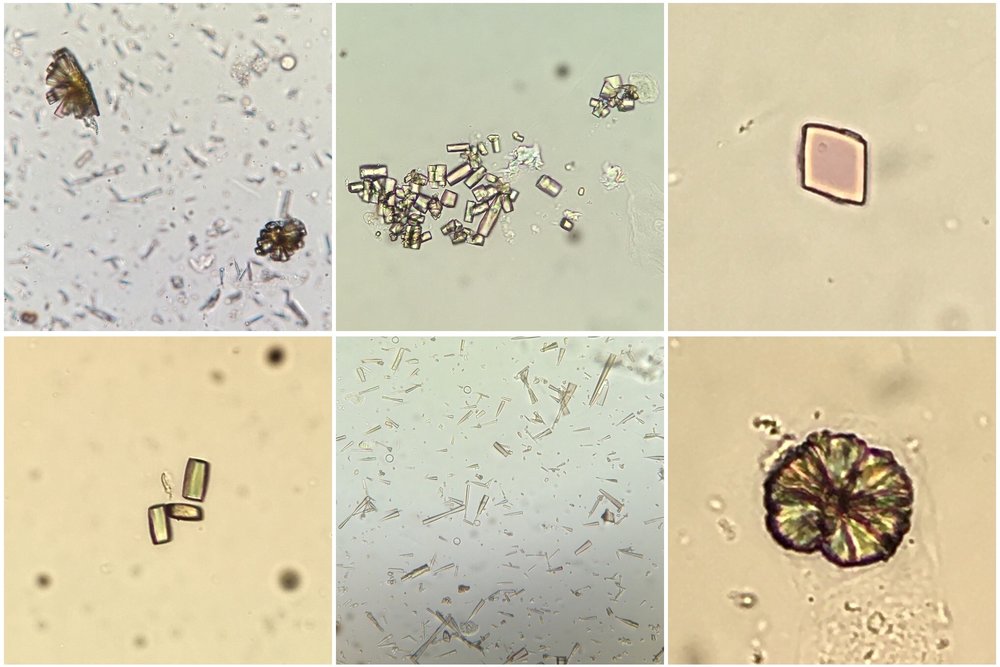
In addition to tumour lysis syndrome, acute urate nephropathy can be caused by other states of tissue catabolism such as seizures, in primary overproduction of uric acid or in cases of reduced urate reabsorption in the proximal tubule. Urinalysis can show uric acid crystals (birefringent with polarisation; see image) or can be normal (as in our patient) perhaps due to a lack of output from obstructed tubules. 
This case raised several points to me. Was his pericardial effusion also caused by a urate infiltration?  No clear cause was ever identified at the time and he did not appear ‘uremic’ despite his renal dysfunction.  Could any of this have been prevented if treatment for his hyperuricemia had been commenced earlier?  I also learned:
  • The nuances of spontaneous tumor lysis syndrome (often phosphate & K not hugely elevated).
  • Rasburicase is contraindicated if glucose-6-phosphatase deficient (approximately 20% of Africans).
  • The ‘undetectable’ result of urate after rasburicase administration appears to be due to in vitro activity of the drug in the blood bottle.

Image thanks to Florian Buchkremer @swissnephro

Post by Ailish Nimmo

Thursday, February 9, 2017

Should you use intravenous immunoglobulin (IVIG) in the treatment of BK nephropathy?

Nice debate on ASN Transplant Community about the use of IVIG for BK nephropathy.

Personally, I have used IVIG in a handful of patients with refractory BK nephropathy in the past. However, I have convinced myself that there is no data supporting that.

Most case series used IVIG in combination with reduction of immunosuppression (Sener et al. Transp 2006), preventing any conclusion on the matter. We had a nice debate with Jay Fishman (MGH Transplant Infectious Disease expert) on last AST Fellow's Symposium about this issue. 

The conclusion was that an effective viral response requires cytotoxic T cells to kill infected cells (Figure above). Antibodies against the virus may help neutralize circulating virus but alone are not capable of stopping an ongoing infection. In particular, prior immunity against BK (IgG BK positive prior to transplant) does not seem to protect against post-transplant BK infection (different than CMV exposure). However, one must keep in mind that there are at least 4 different BK genotypes and immunity against one genotype does not equal immunity to all genotypes, which may explain some of the controversies on the topic (Pastrana et al. PLOS Pathogens 2012).
Overall, reduction in immunosuppression remains the cornerstone of BK viremia treatment.
Remaining controversial topics: 
  • Should you give steroids if intense inflammation? Some data suggest it may not be good... 
  •  Should you stop or just reduce antiproliferative dose? Unclear on my view, but intensity of viremia and allo-immune risk must be balanced here. 
  • Should you switch to an mTOR inhibitor? Some interesting data suggesting that mTOR inhibitors suppress BK replication (similar to suppression to other virus like CMV) (Hirsch et al. AJT 2016)
More data still needed... 

Thursday, February 2, 2017

Cidofovir nephrotoxicity and Probenecid

I recently saw an interesting case. A woman was being treated with cidofovir for adenovirus which was presumed to be responsible for an acute cardiomyopathy. Concurrent with cidofovir, she was also receiving probenecid for renoprotection, which I was not familiar with.
Cidofovir is a nucleotide analogue used primarily to treat CMV retinitis in patients with AIDS. However, cidofovir is also used to treat a number of DNA viruses including adenovirus. The main toxicity of cidofovir is nephrotoxicity, which can manifest as AKI, proteinuria, or a Fanconi-type syndrome with proximal tubular dysfunction. Nephrotoxicity can be reduced by co-administration with iv fluids and probenecid (the dosing regimen for the latter is 2g po 3 hours prior to the dose, then 1 g po 2 hours and 8 hours after.
How does probenecid reduce cidofovir nephrotoxicity? Over 80% of cidofovir is excreted unchanged in the urine in 24 hours. Most of this occurs via glomerular filtration, but cidofovir is also actively taken up from blood by the kidneys via the "organic anion transporter" located on the basolateral side of renal proximal tubular cells, and is then more slowly secreted into the tubular lumen. Renal clearance of cidofovir therefore exceeds the corresponding GFR.
The relatively slow secretion of cidofovir into the tubular lumen, in comparison to uptake from the blood, results in a long intracellular half-life of the drug in the proximal tubular cells which appears to underlie the nephrotoxicity. Probenecid, by inhibiting the organic anion transporter, prevents tubular uptake and protects the kidneys. This was demonstrated nicely in a pilot study in HIV patients. Interestingly, and somewhat paradoxically, this means that probenecid reduces nephrotoxicity while also DECREASING the renal clearance of the drug and thus INCREASING serum cidofovir concentrations as much as two-fold.
Probenecid is a banned drug for athletes for a related reason - because it blocks entry of certain drugs into the urine, it has been used as a masking agent for other banned performance-enhancing drugs including steroids. 
Posted by David Leaf

Wednesday, February 1, 2017

Hyponatremia and ESRD

Hyponatremia can be seen in patients with end-stage renal disease (ESRD), often as a consequence of a patient’s increase in free water intake in the setting of the kidneys’ diminished ability to regulate sodium and water homeostasis. I recently received a question from a resident asking for some insight into the management of such patients.
To begin, it should be noted that uremic patients with chronic hyponatremia are thought to be protected from osmotic demyelination syndrome (ODS) after hemodialysis. In this situation, urea may act as an effective osmole, whereby the decline in blood urea nitrogen (BUN) levels during dialysis could offset serum hypertonicity.  Additionally, animal studies have shown that azotemic rats were protected from ODS due to a reaccumulation of organic osmolytes  such as myoinositol and taurine within two hours of correction of hyponatremia
Though rare, case reports of ODS in uremic patients with hyponatremia do exist. One case described a uremic patient who developed ODS after being initiated on hemodialysis with an initial serum Na of 100 meq/L that corrected to 121 meq/L after three hours against a dialysate sodium concentration of 140 meq/L. It is important to note that ESRD patients in other case reports who went on to develop ODS had other risk factors for the development of the disease, such malnutrition and chronic alcoholism.
I recently helped care for an ESRD patient who was admitted with probable sepsis. His serum sodium on admission was 122 meq/L.  Because we had no outside records available at the time, we assumed his hyponatremia had been present for at least 48 hours. The patient had no uremic symptoms and had no need for urgent small solute clearance or volume removal. Nephrology was consulted for routine dialysis needs.
In choosing our dialysis prescription, we attempted to limit the increase in sodium to no more than 1 meq/L/h, or 3 meq for a 3-hour treatment.  We elected to reduce the blood flow rate to 100 ml/min, use the lowest possible sodium concentration bath allowed in the dialysate (130 meq/L), and use a dialysate flow rate of 600 mL/min.  Cocurrent flows were not used.  After a 3-hour dialysis session, the patient’s serum sodium rose to 125 meq/L, and hourly measurements of serum sodium during dialysis revealed that the patient’s sodium had indeed risen by 1 meq/L/h.
Our calculation was derived from a helpful approach described by Wendland and Kaplan, which showed that the rate in rise of serum sodium could be estimated by the product of the concentration gradient between the patient’s sodium and the dialysate sodium multiplied by the clearance. To do this, we would begins with the following formula:
Change in total body Na = Clearance (L/h) * (dialysate Na – patient Na)
By minimizing the blood flow rate to 100 mL/min and maximizing the dialysate flow rate to 600 mL/min, we assumed that there would be near-total equilibration between the patient’s serum sodium and the sodium concentration of the dialysate bath.  Thus, clearance would be approximately equal to the blood flow rate.
Thus, using our patient as an example:
Change in total body Na = Blood flow rate (L/h) * (130 meq/L – 122 meq/L)
A blood flow rate of 100 mL/min = 6 L/h
Therefore, the change in total body Na after 1 hour of dialysis would be:
6 L/h * 8 meq/L = 48 meq/h
Our patient’s total body water was 48L.  If we add 48 meq Na to our patient’s initial total body Na of 5856 meq (48L * 122 meq/L), we obtain the new total body Na after 1 hour of dialysis: 5856 meq + 48 meq = 5904 meq.
Dividing this value by the patient’s total body water would give us the new serum sodium after 1 hour of dialysis, assuming no ultrafiltration or volume changes.
5904 meq/48L = 123 meq/L
Thus, the patient’s serum sodium would rise by approximately 1 meq/L after 1 hour.
It would be interesting to discuss other options are available for managing ESRD patients with chronic hyponatremia.  Assuming there is no urgent need for small solute clearance, would continuous renal replacement therapy be a safer option?  If uremia is indeed “protective” against ODS, should we avoid low dialysate flow rates and concurrent flows?  How would the prescription change if the patient were to need more aggressive small solute clearance?
It seems that there is more than one way to manage these patients, and factors such as co-morbidities and electrolyte abnormalities need to be taken into consideration when formulating a dialysis prescription.
Posted by Devika Nair, MD
Nephrology Fellow, Vanderbilt