Showing posts with label renal imaging. Show all posts

Tuesday, February 21, 2017

Angiomyolipomas

I got an econsult this morning on a young patient with an incidentally-discovered lesion on her kidney which was consistent with an angiomyolipoma. It is not uncommon to see these on ultrasound and there are a few clinical pearls that I thought I would share:

  • Angiomyolipomas (AML) are benign tumors comprised of blood vessels, adipose tissue and smooth muscle. Very occasionally (more commonly in patients with tuberous sclerosis complex), they can be malignant. Malignant AMLs are more likely to be fat-poor, hypoechoic tumors (termed epitheliod AML).
  • The prevalence is about 2% in the general population and they are 4 times more common in women
  • The presence of multiple AMLs suggests the possibility of tuberous sclerosis complex and the possibility that patients have subclinical disease should be kept in the back of your mind.
  • Confirmation of the ultrasound findings should be done with CT or MRI as there are some renal cancers that can be hyperechoic on ultrasound.
  • The hyperechoic nature of these lesions on ultrasound is due to fat in the tumor rather than the vascularity.
  • For unkown reasons, more of these tumors are found on the right side
  • Most of these tumors are slow growing and do not require treatment (surgery or embolization) unless they are >4cm in diameter. mTOR inhibitors may be useful in some patients with unresectable tumors. Patients should be followed-up yearly with ultrasound.
  • Patients on estrogen therapy are more likely to have rapid tumor growth and should be followed up more often.
Image from: http://www.scielo.br/scielo.php?script=sci_arttext&pid=S0100-72032014000800377

Wednesday, May 4, 2016

Emphysematous Pyelonephritis


http://www.indianjnephrol.org/articles/2013/23/2/images/IndianJNephrol_2013_23_2_119_109418_f6.jpgI was asked to see 74 year old man with an acute on chronic kidney injury.  He had 2 days of generalised lower abdominal pain and vomiting but no urinary symptoms or fever.  His past history included advanced CKD, benign prostatic hypertrophy and a slow growing renal cell tumour under radiological surveillance.  His vital signs were normal and he had mildly raised inflammatory markers.  I ordered a CT KUB to exclude obstruction (it was a weekend and was no ultrasound service in the hospital).  To my surprise this came back as showing emphysematous pyelitis.  Interestingly there had been a hiss of air as he was catheterised for fluid balance monitoring – a fact I had dismissed at the time!


Emphysematous UTIs are gas forming infections of the urinary tract and can manifest as cystitis (gas within the bladder wall), pyelitis (gas within the collecting system) or pyelonephritis (gas within renal parenchyma or perinephric tissues).  It is a relatively rare condition and there is a dearth of literature describing incidence.  Diabetes and urinary tract obstruction are major risk factors, present in around 80% and 20% of patients respectively. Causative organisms are most commonly E. Coli and Klebsiella pneumoniae, with Candida being involved less frequently. Presentation is usually similar to acute severe pyelonephritis with fever, flank pain and vomiting. 50% of patients have an associated bacteraemia. Diagnosis is usually made by CT which shows the extent of gas within the urinary tract and any obstruction.
Treatment depends on the extent of infection.  It ranges from parenteral antibiotics alone for patients where gas is limited to the collecting system with no obstruction, to percutaneous drainage of purulent material and antibiotics if there is abscess formation or extension of gas into the perinephric space, to nephrectomy in patients with diffuse gas and extensive renal destruction.

In the above case, the urine sample was initially reported as ‘no significant bacteriuria’ but subsequently grew a resistant E. Coli >1,000 - <10,000 cfu/ml.  Urology felt that surgical intervention was not required as the renal parenchyma was not involved and he had no abscess formation.  The patient completed 2 weeks of ertapenem and his renal function returned to baseline.
I took several learning points away from this case:
  • As someone who spends a lot of time signing off patients’ results, I realise that ‘no significant bacteriuria’ is not the same as ‘no growth’, and in this case the difference was substantial.  The wording of how we report things and how we interpret that is crucial.
  • A high index of suspicion is required to diagnose emphysematous UTIs and the most appropriate imaging modality should be considered.  Ultrasound is generally the first line investigation for urinary obstruction in patients with acute kidney injury or febrile urinary tract infection due to high sensitivity for hydronephrosis, lack of ionising radiation and lower cost than CT. Ultrasound appearances in emphysematous UTIs can be difficult to interpret however: gas, calculi and calcifications are hard to distinguish and there is often variability in how they are reported. CT is able to precisely localise the presence of gas within the urinary tract and determine whether there is involvement of the renal parenchyma and perinephric tissues.  It can also identify any concomitant pathology or alternate diagnosis e.g. renal calculi.  CT is therefore preferable for diagnosis and subsequent severity staging.
  • Pneumaturia has been described as a presenting feature of emphysematous UTI.  Other causes include vesicovaginal or vesicoenteric fistulae, renal tumour infarction and recent instrumentation.  The unexpected air hiss when catheterising this gentleman was a warning of a more serious pathology and should prompt further investigation.

While the outcome of emphysematous pyelitis is better than that of pyelonephritis (which has a mortality of 18-70% depending on extent of involvement), it is still not a condition to be taken lightly. 
Post by Ailish Nimmo, Royal Infirmary of Edinburgh

Wednesday, March 9, 2016

The evolving role of PET imaging in Nephrology

Positron emission tomography (PET) is a nuclear imaging modality that provides functional imaging of structures based on their ability to metabolize glucose and concentrate specific molecules that have been labeled with a positron-emitting radionuclide. Metabolically active cells (e.g, malignant or inflammatory) utilize and import more glucose than other tissues and, thus, take up 18F-fluorodeoxyglucose (18F-FDG) more avidly. Integrated PET/CT is preferred, because it allows for more exact anatomic localization of isotope uptake and more accurate staging of cancers. PET is escaping oncology however and starting to find a role in nephrology.

In post-transplant lymphproliferative disorders, integrated PET/CT is used as a measure of disease activity. It has also been reported to differentiate fat-poor angiomyolipomas from renal cell carcinoma with 94% sensitivity and 98% specificity. Integrated PET/CT scanning may also be of utility in differentiating benign versus malignant fractures and highlighting impending ones in patients with multiple myeloma.

What about APKD? Cyst infection is common and may be difficult to diagnose in the presence of sterile urine. Bobot et al compared PET-CT to CT and MRI for a diagnosis of cyst infection. Cyst wall hypermetabolism was considered as a positive PET-CT result. A diagnosis of cyst infection was made in 18 of 32 cases: 14 with positive PET-CT findings, and 4 false negatives. There were no false positives and no hypermetabolism of cyst walls in 9 ADPKD control patients. PET-CT had a sensitivity of 77%, a specificity of 100%, and a negative predictive value of 77% compared to CT alone which had a sensitivity of 7% and a negative predictive value of 35%. Radiation doses were comparable and injection of nephrotoxic contrast was avoided in the former.

Similarly PET-CT may be useful the detection of vasculitis in the large arteries with 29/35 patients with known giant cell arthritis showing active arterial inflammation (sensitivity >80%) . Patients suffering from granulomatosis polyangiitis (GPA) show marked aortic FDG uptake although it is unclear whether this is indicative of atherosclerosis or large vessel involvement. If it is indeed the latter this calls into question the traditional classification of vasculitis. FDG-PET/CT accurately identified organ localizations in 16 patients with GPA, other than in nervous system, eye and skin, but may not bring additional benefit to the usual organ screening.
Retroperitoneal fibrosis is a rare fibro-inflammatory disorder that is most commonly idiopathic (>75%) and part of the IgG4-disease related spectrum. PET-CT has emerged as a useful tool for the assessment of disease activity and also detects any post-treatment residual disease. It may lead to early diagnosis of relapses and may also detect diseased sites other than the peri-aortoiliac tissue.

Could PET imaging have a role in the detection of occult malignancy, inflammation or infection in secondary glomerulonephritis? If we extrapolate from studies involving venous thromboembolism (VTE), a prospective cohort of 99 patients with a first episode of VTE reported occult cancer identified by PET/CT in 23% of cases with a sensitivity and negative predictive value of 77% and 97% respectively. However this sensitivity is still too low to justify its use as a widespread screening tool in this capacity as nearly 1/4 cancers may be missed.

Patients with ESRD undergoing maintenance hemodialysis are highly susceptible to infections. Of 104 study patients, 73 (70.2%) had positive 18F-FDG PET/CT findings, and a total of 95 major infection foci were identified. 7 (53.8%) of the 13 patients with primary vascular access-related infections had concurrent metastatic foci. 28 patients (26.9%) had their treatments modified by PET/CT results. In this population, positive PET/CT findings led to a significant change in clinical management and independently predicted mortality.

PET/CT may also help non-invasively prevent avoidable transplant biopsies in kidney transplant recipients with suspected antibody-mediated rejection. In 31 transplant recipients, PET/CT was performed in those who underwent biopsy with a positive correlation between mean SUV and acute composite Banff score (r2 =0.49). The area under the receiver operating characteristic curve was 0.93, with 100% sensitivity and 50% specificity using a mean SUV threshold of 1.6.

But are there any side-effects or limitations to use of PET-CT in our patients? For example, use of IV contrast may be precluded in certain cases of renal impairment and this may impair the optimal detection of small lung and liver lesions. IV contrast increases lesion conspicuity, which is of particular importance in the evaluation of lesions that do not always accumulate FDG. However use of PET-CT without IV contrast has become more widespread and differentiation of benign from malignant lesions is less relevant in Nephrology. The whole-body FDG distribution in patients on hemodialysis may be different from those with normal renal function, because they lack urinary FDG excretion and remain in a constant volume overload. There may be significantly higher physiological FDG uptake in the soft tissues, spleen and blood pool.

In conclusion, I believe that combined PET-CT imaging has the potential to be a very useful and versatile tool for Nephrologists. Whether we are dealing with metastatic infections, occult malignancy, suspicious GN or vasculitic relapses, it may be a revealing diagnostic test at times when our concern exceeds the objective evidence that we have to hand. That said, it is not without cost or risk, and therefore should only be employed judiciously when likely to change clinical management.


Post by Dearbhla Kelly,
NSMC Intern

Friday, August 2, 2013

Ultrasound Course for Nephrologists

Continuing our practice of highlighting excellent courses for Nephrologists and Fellows, here is a message from Professor W. Charles O'Neill regarding the annual Ultrasound for Nephrologists Course at Emory University this October. The course brochure and application form are here

"Ultrasonography is an invaluable tool in nephrology and, following the lead of other specialists, nephrologists are beginning to incorporate ultrasound into their practice. It is convenient, economically feasible and improves patient care. Since this skill is not generally taught in training programs, we have been offering the Ultrasonography for Nephrologists course. The course has been very well received and has been given continuously since 1997. It is a comprehensive, weekend program that provides both didactic and hands-on training in ultrasonography of native and transplanted kidneys, end-stage kidneys and allografts, and guidance for biopsy and central venous catheterization. There are 6 hours of hands-on training that include patients with a variety of abnormalities. Also covered are financial aspects including equipment costs, billing, reimbursement, and other aspects such as reports, record-keeping, databases, quality assurance, and accreditation. There will also be an opportunity to use equipment from a variety of manufacturers. Pretty much everything a nephrologist needs to get started."

Friday, October 5, 2012

Less is More


Despite the fact that the kidney ultrasound is generally obtained as one of multiple recommendations when evaluating AKI, the benefit of kidney ultrasound is not clear. The post-renal causes of AKI are not very common. It adds to the cost and can subject patients to unnecessary work-ups by revealing incidentalomas. How often, then, is the ultrasound useful? 
In order to assess the prevalence of the post-renal AKI and determine a cost-effective use of the ultrasound, a single-center case-control study was conducted.
Their conclusion is that the prevalence of hydronephrosis requiring stenting or nephrostomy placement was only 0.4% in the low-risk group. The number to screen to find a case of urinary obstruction was 223. At what cost? In our institution the kidney ultrasound without Doppler costs $600. It costs $133,800 to find one case.
Who is the low-risk group patient? Based on the multivariate analysis, a patient was considered low-risk if he or she did not have a history of hydronephrosis and had no more than one of the following:
1. Recurrent UTI
2. Diagnosis to suspected obstruction (BPH, abdominal or pelvic cancer, one functional kidney, neurogenic bladder, pelvic surgery)
3. Non-African American
4. Absence of:  exposure to the following medications (ASA, diuretics, ACEI or IV vancomycin), congestive heart failure, or pre-renal AKI.
The study has limitations. Not all AKI patients were studied. The cases requiring non-surgical interventions were not counted. If we would have to implement this strategy, we don’t know what the cost of missing some cases of obstruction would be.
However, the implication is that we should not routinely order kidney ultrasound on every patient with AKI, particularly those in the low-risk group. In this era of cost constraint on medicine, less is usually more…
Or, here is what you can do. If your place has an ultrasound on the floor, with a little training you can have a quick look at the kidneys just to rule out obstruction in low risk patients. You acquire one more diagnostic skill, your students have one more fun on round, and your hospital saves significant amount of money!
Posted by Tomoki Tsukahara

Sunday, May 6, 2012

Diagnosis and Management of Post-Transplant Fluid Collections

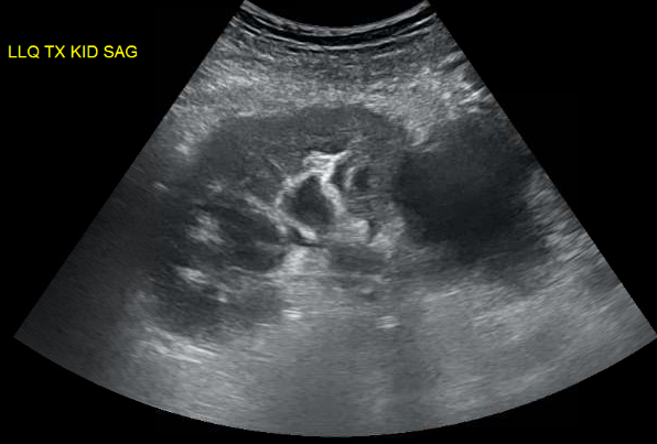
We were evaluating a kidney transplant recipient three weeks after surgery who presented with a rise in creatinine. We obtained an ultrasound which showed a large fluid collection and mild hydronephrosis. Prograf level was at goal. 
Peritransplant fluid collections may be produced by lymphoceles, urinomas, hematomas or abcesses. These collections may compress the ureter leading to hydronephrosis or may compress the renal vein causing a “compartment-like syndrome”. Since his creatinine was risen, we decided to drain the fluid collection. An important point is that mild hydronephrosis is reported in a large portion of kidney transplant ultrasounds and intervention is usually driven by the clinical setting. When in doubt, serial ultrasounds indicating progressive dilatation or the use of nuclear medicine imaging showing normal perfusion and parenchymal uptake but pooling of tracer in the renal pelvis with prolonged pelvic retention helps in confirming the diagnosis. 
His INR was 2.1 due to coumadin for a history of multiple clotted fistula. The interventional radiologist requested that we give FFP to reduce his INR to below 1.5 before considering the procedure. It was late Friday so we got 4U of FFP in and repeated INR. We were hoping that 4U of FFP would quickly reverse his mildly elevated INR. Interestingly, data supporting that is lacking. 
FFP can have an INR as high as 1.5 and transfusion of FFP will have little effect on minimally elevated INRs (Gearoid actually pointed that out in rounds). A study of the use of FFP in 121 patients with an INR in the range of 1.1 to 1.85, showed that correction of the INR to normal was achieved in only one patient (0.8 percent) and correction at least halfway to normal in only 15 percent. The median decrease in the PT and INR was 0.2 seconds and 0.07, respectively, and was independent of the number of units of FFP infused (median 2 units; range 1 to 20). Thus, available studies do not support the efficacy of FFP in treating bleeding or as prophylaxis for invasive procedures in patients with a mild coagulopathy (ie, INR below 2.0). 
After some discussion with IR, they agreed to go ahead with the procedure with an INR 1.9. About 600cc of yellow fluid was drained. Fluid creatinine was checked and was similar to serum level, ruling out an urinoma. This patient’s fluid collection was a lymphocele, which is the most common cause of peritransplant fluid collection due to disruption of renal lymphatics. It usually occurs weeks after surgery and drainage is required only in cases of suspected obstruction. With time, the amount of fluid collection tends to subside. Soon after drainage, his creatinine started to come down confirming the cause of his worsening renal function.

Figure: large peri-transplant fluid collection with associated hydronephrosis.

Thursday, June 17, 2010

Evaluating incidental renal cysts

Last week, I got an email from a residency friend asking me what I would do with a patient who had a Bosniak II renal cyst. Since “I don’t know” no longer seemed a satisfactory answer for someone who specializes in kidneys, I looked it up, returned her email, then decided to review the radiologic classification of renal cysts on the blog, for anyone else who would like a refresher. An excellent review by Glickman et al can be found in the journal Radiology.

Renal cysts are masses filled with fluid. They are extremely common: some studies have estimated their incidence in the general public at around 50%. Most are discovered incidentally, since they rarely cause symptoms. Flank pain and hematuria are occasionally seen. Radiologic features of renal cysts convey important information about their malignant potential—our main concern as physicians. Does the wall of the cyst appear thin and simple, or does it have a thickened lining? Is the fluid in the cyst attenuated? How heterogeneous does the cyst appear? Are there septae or enhancing soft tissue components? In 1986, a classification scheme was published by Bosniak that stratified malignant risk based on characteristics of the cyst on CT scan. The Bosniak classification remains a useful guide for management of incidentally-discovered renal cysts. It should be emphasized, however, that evaluation of cysts must take into account a patient’s demographics and medical history: a diagnosis cannot be made by imaging alone.

Bosniak Classification of Renal Cysts

Bosniak I- benign, simple cysts, with thin walls. Note that size is not a factor in classifying cysts in this category. Bosniak I cysts are always benign, and can be ignored. These cysts are not attenuated, and tend to have Hounsfield units of 0-20.

Bosniak II- benign, minimally complicated cysts; may have hairline-thin septa with some perceived enhancement. There may be fine calcification or areas of calcified thickening along the wall or septa. Fine calcification or a short segment of slightly thickened calcification may be present in the wall or septa. Homogeneous hyperattenuating cysts also belong in this category—high Hounsfield units usually signify proteinaceous fluid. Bosniak II cysts are also felt to have non-malignant potential, and can be ignored by the physician.

Bosniak IIF- these are slightly more complicated that class II cysts, and so warrant observation. IIF cyts may contain multiple, hairline-thin septa that demonstrate perceived (but not measurable) enhancement. There may be minimal smooth thickening of the wall or septa. Calcification that is more irregular and thicker than in Bosniak II cysts may be present. Soft tissue elements are absent. Hyperattenuating cysts otherwise classified as category II cysts, but are > 3cm and completely within the renal parenchyma are included in class IIF. The general recommendation for observation of these cysts is to reimage with CT or MR in six months, then yearly for a minimum of five years. If they remain stable, it can be assumed that they are benign, and no further follow-up is needed.

Bosniak III- the malignant potential of class III masses is indeterminate on imaging; therefore, surgical removal is recommended. These cysts have thickened walls or septa, and display enhancement. They include multilocular cysts (in which the walls have fibrous lining), hemorrhagic or infected cysts, multilocular cystic nephroma (which conatin blastemal elements), or cystic renal cell carcinoma. It is estimated that 50% are malignant (studies have shown malignancy rates ranging from 31% to 100%).

Bosniak IV- share features of Bosniak III, plus enhancing soft-tissue components adjacent to or separate from the wall or septa. They are almost always malignant. Management is surgical.


The Bottom Line: radiologic followup is needed only for class IIF lesions. The others are either ignored or referred for surgical management secondary to the high rates of malignancy.

Tuesday, July 21, 2009

Caveats for Using Renal Ultrasound to Diagnose Post-Renal Failure

Most would agree that the renal ultrasound is an invaluable tool in the workup of acute kidney injury. Although the renal ultrasound can give information regarding kidney echogenicity and presence of renal cysts, by and large its predominant utility is in determining the presence or absence of hydronephrosis, a marker for obstructive renal failure ("post-renal" failure when referring to the Holy Trinity of "pre-renal", "intrinsic renal", and "post-renal" categories of AKI).  

However, there are important caveats to the use of the renal ultrasound to effectively rule-in or rule-out obstructive nephropathy.  For instance, false-negatives (e.g., the renal ultrasound does not show hydronephrosis, but there actually is) can occur in the following scenarios:

1.  very early obstruction:  truly acute obstruction (e.g., with a kidney stone, for instance) may take some time to develop enough distension of the collecting system to detect with our current imaging tools.
2.  obstruction in the setting of pre-renal failure:  volume-depleted kidneys may not demonstrate significant hydronephrosis until after volume resuscitation, which expands the collecting system.
3.  large retroperitoneal tumors:  tumors which encase the kidneys may cause obstruction but not allow expansion of the collecting system enough to see hydronephrosis.  
4.  retroperitoneal fibrosis:  this can occur in patients with past extensive GU surgery or prior chemotherapy or radiation therapy and can cause mechanical obstruction without allowing expansion of the collecting system.

In addition, false-positives (e.g., the renal ultrasound is read as "hydronephrosis" but this is probably not pathologic) can occur as well, most notably in pregnancy.  It is also common that hydronephrosis detected on ultrasound is a chronic, long-standing issue and not the main culprit for the present episode of AKI. 

This post is summarized from some basic but well-written handouts by Dr. Joel Topf from Precious Bodily Fluids.  Not a bad idea to print these out and go over cases with a medicine resident rotating on the nephrology consult service...

Wednesday, October 22, 2008

Differential Diagnosis for Large Kidneys

The differential diagnosis for large kidneys (often demonstrated on renal ultrasound) is interesting, as there are a few conditions which are especially associated with this finding. Of course, with any type of chronic damage, the kidneys tend to scar down and atrophy.

1. HIV Nephropathy.
2. multiple myeloma.
3. the early stages of diabetic nephropathy.
4. any type of infiltrative process--classically amyloidosis or sarcoidosis of the kidney.
5. autosomal dominant polycystic kidney disease.
6. pyelonephritis.
7. any type of very acute nephritis (e.g., RPGN).

Sunday, August 3, 2008

Urinoma

Time for some "Renal Radiology Rounds."

The case involves a 77 year-old man with a history of severe benign prostatic hypertrophy. He had been advised to have a TURP in the past but had repeatedly declined. He was hospitalized for nausea, vomiting and R flank pain without hematuria occurring over a week's time, and it was noted that his creatinine had elevated from its baseline of 1.3 to 7. A CT scan on arrival showed the following:


Uploaded on authorSTREAM by nathanhellman

This patient had developed a R-sided urinoma, which is simply a collection of urine which results from rupture of the collecting system due to high pressures from obstruction. It is more typically seen in renal transplant patients post-operatively at the site of the surgical anastamosis between the recipient and donor collecting systems; non-surgical cases of urinomas such as this one are more rare. The patient had a decompressive Foley catheter placed and an IR drain placed to remove the fluid collection. The fluid's creatinine level was higher than serum level, indicating urine as the source. He experienced a post-obstructive diuresis with a lowering of his creatinine following these interventions.

Thursday, July 10, 2008

What is the Resistive Index?

Often on the official radiology report for a renal ultrasound with Doppler flows, a number called the "resistive index" is listed.

What does this number mean and how is it derived?

The resistive index (RI) measures the resistance of renal arterial flow to the kidney. In a normal situation, flow through the renal artery occurs throughout systole and diastole. However during a variety of conditions--transplant rejection, inflammation, obstruction--there will be reduced to possibly even reversed flow through the renal artery during diastole. When this happens, the resistive index--which is calculated by measuring the arterial waveform throughout the cardiac cycle as described in the figure--is elevated. An RI <0.8>0.9 is considered elevated.

The differential diagnosis for an elevated RI should include transplant rejection, obstruction, ATN, pyelonephritis, severe hypotension, or an acute vascular event (e.g. renal vein thrombosis).

Monday, July 7, 2008

Nuclear Medicine Renal Imaging

There are numerous different types of radionuclide imaging which are used in renal imaging--more commonly in pediatric than in adult patients, and as such my knowledge of these procedures as it pertains to adult nephrology patients has always been a bit hazy.

Fortunately there is an excellent review of this topic in this month's Nephsap from ASN featuring Renal Imaging.

One of the tests often ordered in pediatric populations is that so-called "Mag-3 Renal Scan". This stands for technitium-99m-mercapto acetyl tri glycine-3, the radiotracer used in this form of imaging termed "dynamic renal scintigraphy." The radiotracer is injected intravenously and then dynamic images of the collecting system are obtained over a 30-minute period, over which time the tracer should be excreted and appear in the bladder. This technique is useful for demonstrating functional obstruction--for example, due to a kidney stone or other anatomic abnormality--based on the rate of Mag-3 washout from the collecting system, particularly in instances when other imaging modalities (e.g., ultrasound, CT urography) fail to demonstrate a definitive obstruction but there is still a high suspicion that one exists.

Other commonly used nuclear medicine renal imaging tests include "renal cortical scintigraphy" (in which the radiotracer Tc-99m-DMSA is used to demonstrate acute or chronic pyelonephritis/scarring, usually in pediatric populations) and "radionuclide cytography" (which assesses for vesico-urethral reflux by injection of a technicium radiotracer in either an anterograde or retrograde fashion.)

Wednesday, April 23, 2008

Ultrasound Criteria for ADPKD

Autosomal dominant polycystic kidney disease (ADPKD) acconts for about 10% of patients on dialysis in the U.S. and occurs in 1:400 - 1:2000 live births. As the disease is inherited in an autosomal dominant fashion, the question of genetic counseling often comes up (e.g., does my brother/sister/son/daughter also have ADPKD?) This is also a very relevant question in deciding whether or not a relative of a patient with ADPKD is an appropriate kidney donor: obviously, you wouldn't want to take PKD kidney as a transplant, which would have poor results for both the recipient as well as the donor. Unfortunately, mutation screening is not useful in ADPKD since the PKD1 gene (accounting for about 85% of cases of ADPKD) is extremely large with multiple internal repeats, making it difficult to sequence, and furthermore the list of possible ADPKD mutations numbers in the 100s rather than being just a few common mutations.

To this end, a series of ultrasound criteria have been created and validated for screening. The criteria are based on the principle that the younger you are, the less likey you are to have cysts. These criteria have a very high sensitivity & specificity (apparently approaching 100%) in the appropriate clinical scenario. The criteria are as follows:

For patients with PKD 1 mutations:
age <30:>60: need at least 4 cysts per kidney.

For patients with PKD 2 mutations (which is generally less severe and delayed onset compared to PKD 1 mutations:

-need 4 or more cysts per kidney (ages 30-59) to reach a sensitivity of 96%.

According to recommendations on Up To Date, patients less than 18 years of age should NOT be screened for ADPKD.

References:

Ravine, D, Gibson, RN, Walker, RG, et al. Evaluation of ultrasonographic diagnostic criteria for autosomal dominant polycystic kidney disease 1. Lancet 1994; 343:824.

Nicolau, C, Torra, R, Badenas, C, et al. Autosomal dominant polycystic kidney disease types 1 and 2: Assessment of US sensitivity for diagnosis. Radiology 1999; 213:273.