Showing posts with label tumor lysis syndrome. Show all posts

Tuesday, February 21, 2017

Spontaneous Tumor Lysis Syndrome

A 70 year old Ghanaian man was recently admitted under our care.  He had been diagnosed with aggressive myelodysplasia 2 months previously after presenting with fatigue and abnormal blood results (WBC 50.3, platelets 130 and LDH 928 at the time of diagnosis).  A plan was made for palliative chemotherapy. One month after his diagnosis he developed a large pericardial effusion and had 1L of haemorrhagic fluid drained.  At this point his creatinine was 200 umol/L (2.26 mg/dL).  Routine and TB culture of the fluid was negative, as was cytology and immunophenotyping.
Image result for swissnephro uric acid
Two weeks after this admission he represented with abdominal pain.  A CT showed bilateral renal and bladder calculi without obstruction.  He was oliguric with a creatinine of 577 umol/L (6.5 mg/dL) rising to 709 umol/L (8 mg/dL) over the next 12 hours.  His uric acid level was 18.0 mg/dL, which had not been checked previously.  Phosphate was 1.86 mmol/L (5.75 mg/dL), Ca 2.1 mmol/L (8.4 mg/dL) and K 4.6 mmol/L.
Our diagnosis was of a spontaneous tumour lysis syndrome (TLS; see previous RFN posts here & here).  Nucleic acids released from tumour cell lysis are broken down into xanthine and then uric acid by xanthine oxidase.  Renal failure is caused by uric acid precipitating in renal tubules causing a mechanical obstruction and inflammatory reaction.  While TLS is typically seen following initiation of chemotherapy causing a rapid breakdown of cancer cells, a spontaneous form has been described in acute leukaemia and NHL.  Our patient was at high risk of converting into AML but had no rise in peripheral blasts to suggest this.
Interestingly, spontaneous tumour lysis syndrome is associated with hyperuricemia but often without the hyperphosphatemia (and hyperkalemia) seen in the classical form of the disease– thought to be because the released phosphorus is quickly used up in the generation of new tumour cells.  This would fit with our patients results.
Our patient was commenced on dialysis which gave reductions in uric acid levels of 50% per treatment, but they quickly rebounded.  He was no longer fit for treatment of his myelodysplasia making longer term management more difficult.  Given his African ethnicity, we checked his glucose-6-phosphatase levels, which were normal, before he received rasburicase (recombinant urate oxidase). Rasburicase reduces uric acid levels by converting it into allantoin.  It may cause severe oxidative hemolysis if glucose-6-phosphatase deficient. Uric acid fell to undetectable levels following this however he had an ongoing dialysis requirement (note that rasburicase retains in vitro activity in the blood bottle so sample should ideally go on ice).  Allopurinol as a longer term medication to reduce uric acid formation may be useful, but may not manage to suppress formation sufficiently.  
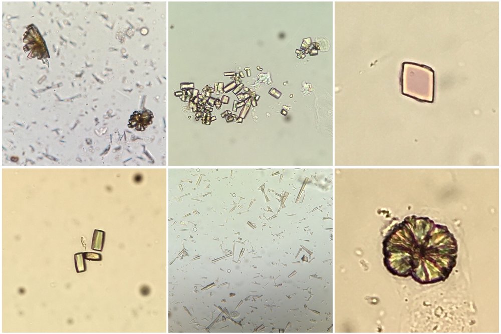
In addition to tumour lysis syndrome, acute urate nephropathy can be caused by other states of tissue catabolism such as seizures, in primary overproduction of uric acid or in cases of reduced urate reabsorption in the proximal tubule. Urinalysis can show uric acid crystals (birefringent with polarisation; see image) or can be normal (as in our patient) perhaps due to a lack of output from obstructed tubules. 
This case raised several points to me. Was his pericardial effusion also caused by a urate infiltration?  No clear cause was ever identified at the time and he did not appear ‘uremic’ despite his renal dysfunction.  Could any of this have been prevented if treatment for his hyperuricemia had been commenced earlier?  I also learned:
  • The nuances of spontaneous tumor lysis syndrome (often phosphate & K not hugely elevated).
  • Rasburicase is contraindicated if glucose-6-phosphatase deficient (approximately 20% of Africans).
  • The ‘undetectable’ result of urate after rasburicase administration appears to be due to in vitro activity of the drug in the blood bottle.

Image thanks to Florian Buchkremer @swissnephro

Post by Ailish Nimmo

Monday, June 25, 2012

To Treat of Not


A 46 year old man with a recent diagnosis of acute leukemia and white count of 90,000 was transferred to our facility with non-oliguric AKI secondary to tumor lysis syndrome. The patient was hypocalcemic (Ca 5.2mg/dl) and hyperphosphatemic (PO4 11 mg/dl). His K was 5.8 mmol/L and his creatinine was 3.5 mg/dl. He was making around 200mls/hr of urine and he was asymptomatic. The Hem/Onc resident called nephrology looking for advice about whether or not the hypercalcemia should be treated and if he would be better off getting D5W with bicarbonate instead of normal saline.
TLS is a result of rapid and massive breakdown of tumor cells, either spontaneously or after the initiation of cytoreductive therapy. Because potassium is stored primarily in the intracellular compartment, its rapid release into the extracellular compartment during TLS can lead to hyperkalemia. Similarly, hyperphosphatemia results from a massive release of intracellularly stored phosphate, that can lead to secondary hypocalcemia. Uric acid is the end product of the catabolism of purines, which are released from the breakdown of nucleic acids. That lead us to the laboratory definition of TLS, that was developed by Cairo and Bishop.


Renal injury can result from two components; the deposition of uric acid and calcium phosphate crystals, and non-crystal mechanisms including renal vasoconstriction, alteration in renal autoregulation through inhibition of nitric oxide synthesis and a resulting decrease in endothelial cell nitric oxide, and stimulation of the renin-angiotensin system.
It is best to avoid IV calcium administration unless hypocalcemia is symptomatic because it might increase the risk of calcium phosphate precipitation and the potential for additional kidney injury. It is also best to monitor serum ionized calcium levels, especially in patients with hypoalbuminemia.
When rasburicase is available, hyperuricemia is seldom an indication for dialysis. Rasburicase is a recombinant urate oxidase that converts uric acid to the more water-soluble product allantoin (which is not dependent on urinary pH for its solubility).
Uric acid solubility is low and increases as urinary pH becomes more alkaline. However, calcium phosphate is more soluble at an acidic pH; therefore, urinary alkalinization may lead to increased calcium-phosphate crystallization and precipitation.
Therefore, especially when rasburicase is available to manage the hyperuricemia, urinary alkalinization should be avoided.
Going back to the patient, neither Ca nor bicarbonate was given. Uric acid was not measurable the next day following the use of rasburicase. PO4 decreased gradually over the next few days, and no dialysis was required during the hospitalization.
Update: As a commenter pointed out below, rasburicase continues to work in vitro unless the sample is immediately placed on ice and the "undetectable" uric acid level may have been artefactual.
Posted by Tarek Alhamad

Monday, October 11, 2010

Tumor lysis syndrome and acute kidney injury

The May issue of AJKD (Vol.55, No.5) had a nice supplement on TLS and AKI. Here is the link.

Tumor lysis syndrome (TLS) describes a condition with significant clinical and lab abnormalities caused by rapid and massive tumor cell death. Occurring either spontaneously or after chemotherapy, TLS is a medical emergency and is associated with significant morbidity and if untreated mortality.

The
risk for patients of developing TLS changes with their type of malignancy:

1. High risk: Burkitt Lymphoma, Lymphoblastic Lymphoma, B-ALL, acute ALL (WBC > 100K), acute AML (>50K monoblastic WBCs)

2. Intermediate risk: Diffuse large B-cell lymphoma, acute ALL (WBC 10-50K), acute AML (WBC 10-50K), CLL (WBC 10-100K, treated with fludarabine), malignancies with rapid proliferation with expected rapid response to therapy

3. Low risk: indolent NHL, acute ALL with WBC <10k>

Definition of TLS
is as follows:

1. Definition of Laboratory TLS (any 2 or more criteria within 3 days before or 7 days after chemotherapy):

- Uric Acid > 8mg/dl or 25% increase from baseline
- Potassium > 5 mEq/L or 25% increase from baseline
- Phosphorus > 6.5 mg/dl (children) or > 4.5mg/dl (adults) or 25% increase from baseline
- Calcium less than 7mg/dl or 25% decrease from baseline

2. Definition of clinical TLS (laboratory TLS plus at least one of the below criteria):
- Creatinine greater than
1.5 value of upper limit of age-adjusted normal range
- Cardiac arrhythmias or sudden death
- Seizure


Acute Kidney Injury in TLS occurs mainly through crystal deposition including
a) uric acid
b) calcium phosphate

Other aggravating factors include:
c) volume depletion / hypotension / CHF
d) extrinsic urinary obstruction
e) pre-existing CKD
f) nephrotoxic medications such as NSAID
g) radiocontrast exposure
h) sepsis

Prevention and treatment of the AKI caused by TLS focuses on reversing above factors, requires volume depletion, and treatment of electrolyte abnormalities (hyperkalemia, hyperphosphatemia). The primary goal is to minimize or treat hyperuricemia by either:

Decreasing production of Uric acid with Allopurinol, which blocks Xanthin oxidase,
and/or
Converting Uric acid into water-soluble Allantoin with Rasburicase (Urate oxidase)

Of these approved treatments for hyperuricemia, the former with allopurinol can lead to Uric acid independent crystal deposition by accumulation of Xanthin products. In addition, elevated urinary Uric acid levels may lead to crystal-independent AKI by by alteration of renal autoregulation and vasoconstriction. Routine urinary alkalinization to prevent Uric acid crystals is controversial since it may increase risk for renal calcium phosphate crystal deposition. Dialysis therapy should be initiated when indicated using standard criteria for AKI.