Monday, July 16, 2012

Hyperammonemia in Myeloma: Dialyze?


 A middle aged man with IgG kappa multiple myeloma previously treated with bortezomib and lenalidomide presented to the hospital with altered mental status. He had completed chemotherapy months prior to presentation. Shortly after being admitted, he progressed to obtundation associated with tachypnea and a profound respiratory alkalosis requiring intubation for airway protection. His initial arterial blood gas at the time of intubation revealed a pH of 7.35, an undetectably low pCO2, and a bicarbonate of 14 (on concurrent labs) with an anion gap of 14. Mechanical ventilation was initiated with a minimal amount of pressure support. All subsequent blood gases demonstrated a pH greater than 7.55 with ongoing respiratory alkalosis. The anion gap normalized. Intensive work-up of the altered mental status resulted in the sole finding of hyperammonemia with a serum ammonia level of 100 umol/L. There was no liver injury evident on labs. Lactulose was initiated to treat the elevation in ammonia with no improvement noted.

We were consulted regarding the possibility of dialysis to correct the hyperammonemia. 

Do we need to correct high ammonia levels?

Hyperammonemia carries a significant morbidity and mortality, and patients frequently require ICU-level care for encephalopathy. With acute presentations of hyperammonemia, levels of ammonia greater than 200 umol/L are associated with cerebral edema and herniation due to cerebral dysautoregulation. In the presence of chronic hyperammonemia, compensatory increases in ammonia metabolism by the muscles and hepatic and splanchnic vascular beds may blunt symptoms. Interestingly, arterial, venous, and brain levels of ammonia typically do not correlate in patients with chronic hyperammonemia but have a better correlation in acute hyperammonemia. In our patient, we did not check arterial ammonia levels to look for a correlation.

Why is our patient hyperammonemic?

Ammonia is produced primarily in the gut as a product of protein breakdown and bacterial metabolism, and it is broken down primarily in the liver. Increased production of ammonia can occur in the presence of protein breakdown from GI bleeding. In patients with liver failure who are already predisposed to having high ammonia levels, GI bleeding is a known risk factor for the precipitation of hepatic hyperammonemic encephalopathy.

Our patient had no laboratory evidence of liver dysfunction. He had recently suffered an episode of GI bleeding from a bleeding mechanical abnormality seen on EGD that was corrected. Perhaps this could have been the inciting factor for the elevated ammonia, but the patient's ammonia levels remained elevated throughout his hospital stay despite termination of the bleeding.

Inborn errors of metabolism can be considered, but most of these present in childhood. However, urea cycle disorders can be unmasked in adulthood by medications, protein intake, and infections. The drugs most likely to be involved include salicylates, valproate, carbamazepine, sulfadiazine, pyrimethamine, glycine, and TPN. None of these were implicated in our patient. Urea-splitting organisms and herpes infection can also raise ammonia levels. Neither were present.

Alas, multiple myeloma can be a cause of hyperammonemia. Rising beta-2 microglobulin levels in the absence of acute kidney injury in our patient suggested worsening myeloma off of chemotherapy.

Hyperammonemic encephalopathy in multiple myeloma

In vitro, myeloma cell lines secrete ammonia into culture medium in greater amounts than other hematological malignant cells. This may be due to excess protein synthesis in myeloma cells. In vivo, the exact mechanism for hyperammonemia in multiple myeloma patients is unknown.

It is important to rule out hypercalcemia and hyperviscosity as causes of altered mental status in patients with multiple myeloma before encephalopathy is attributed to elevated ammonia levels. In our patient, calcium and viscosity levels were checked and found to be normal.

In a published review of 27 cases of hyperammonemic encephalopathy in multiple myeloma patients, depressed levels of consciousness were noted to occur at ammonia levels ranging from 35 to 39,342 umol/L. The degree of ammonia elevation did not appear to correlate with death (if one ignores the outlier of 39,342) but in all cases where the patient's ammonia level did not improve, death was the unfortunate outcome. Presenting symptoms included respiratory alkalosis, asterixis, myoclonus, hallucinations, and hyperdynamic heart failure. Of the 27 reported cases, 4 patients received dialysis (3 HD, 1 PD) and 9 received ammonia-lowering medications such as antibiotics, lactulose, carnitine, and flumazenil. All patients received chemotherapy.

Should we dialyze our patient?

In drug intoxications associated with hyperammonemic encephalopathy, dialysis serves a definitive role in clearing the toxin and correcting the hyperammonemia. However, in cases resulting from malignancy, the answer is less clear.

Of the 4 patients who received dialysis in the study described above, 3 survived. Of the remaining 23 patients who were not dialyzed, 12 survived. Ammonia levels decreased in all 4 patients who received dialysis and in 19/23 patients who did not undergo dialysis. This suggests that dialysis plays perhaps only a minor role in lowering ammonia in this patient population. Given the potential for harm associated with dialysis catheter placement, we felt that the risks outweighed the unclear (if any) benefits of dialysis.

Our patient received steroids and further chemotherapy. Interestingly, administration of steroids can raise ammonia levels in the short term because of increased catabolism. Ammonia levels fluctuated wildly but improved slightly. The patient was extubated succesfully. His mental status improved but not to baseline. He continued to have a respiratory alkalosis at the time of discharge. 

Ammonia: Chicken, Egg, or Bystander? 

In hepatic encephalopathy, it is well known that ammonia levels do not correlate with the degree of encephalopathy. For this reason, following ammonia levels after initiating treatment is discouraged. The same may be true here. Perhaps ammonia is the cause of the encephalopathy; perhaps it is a biomarker of disease; perhaps it is an innocent bystander. Further study is needed to elucidate this as it may clarify the role of dialysis as a treatment for hyperammonemia in myeloma.

References


Friday, July 13, 2012

Venus, Sjogren's and the Kidney

The Williams sisters put yet another Wimbledon doubles tittle into their trophy case over the weekend.  This, just hours after Serena had won the singles competition.  Pretty impressive.

In reading the coverage I learned that during the last year Venus had been diagnosed with Sjogren's syndrome (I know, I know, old news to those paying attention).  There was no mention in any of the reports that Venus has had any kidney problems, her main issues have apparently been with severe fatigue and joint pains.  Although we usually think of dryness in the eyes and mouth when we hear Sjogren's syndrome there are a number of potential renal tip offs to the diagnosis...

Interstitial (and sometimes glomerular) Disease

Interstitial nephritis - Typically mild but can progress to ESRD in rare cases.  In a Mayo Clinic case series 46% and 25% of patients with Sjogren's syndrome who underwent kidney biopsy had chronic or acute interstitial nephritis respectively.  Only one of the 24 patients biopsied had ESRD and this was from a total case series of 7276 patients (there may have been some additional cases of ESRD lurking in the cohort who didn't get biopsied as they didn't have complete clinical data on all the patients).

Glomerular disease - Less common than interstitial disease but does occur. Associations with MPGN (often in association with cryglobulins), FSGS, Membranous and minimal change have all been reported. 

Tubular defects

Distal renal tubular acidosis - An unexplained distal RTA is a invitation to investigate for Sjogren's.  Patients with distal RTAs are prone to nephrolithiasis (typically calcium phosphate stones) and nephrocalcinosis.  Severe associated hypokalemia can also occur with flaccid paralysis having been described as a presenting symptom.

Although the cause of distal RTAs in most patients with Sjogren's syndrome is unknown a few patients have been described who have an absence of the H-ATPase pump on intercalated cells in the collecting duct.  Another hypothesis is that Sjogren's leads to autoantibodies directed against carbonic anhydrase II thus leading to the generation of less protons for excretion.

Proximal RTA and full blown Fanconi's syndrome have also been described.

Nephrogenic diabetes insipidus - Urinary concentrating defects are not uncommon in patients with Sjogren's.  As an example, in an Italian series 21% of patients were noted to have an abnormal urinary concentrating ability.  The cause of this tubular defect is again unknown but histologically clusters of lymphocytic infiltrate can sometimes be seen around collecting ducts which could theoretically interfere with the actions of ADH.

Hypokalemia - This can occur in the absence of RTA and is thought to be due to tubular damage induced sodium wasting with subsequent increased distal sodium delivery.  In the distal nephron increased sodium delivery drives potassium loss in exchange for sodium.  This effect may be amplified by volume depletion with subsequent increased aldosterone levels which again drive sodium absorption and potassium loss.

Thursday, July 12, 2012

Urea and Hyponatremia


Urea is a hyponatremia treatment long forgotten in the United States. Chronic SIADH is usually managed either by vaptans or a combination of fluid restriction, salt and furosemide. However, vaptans are very expensive and few patients can afford it. In a recent Belgian article, the use of urea as a comparable, cheaper alternative is being advocated.

What do we know about urea? It is a very cheap powder (< $0.50/30 gram) that works by increasing free water excretion through osmotic diuresis. Shown below is the solute excretion as a determinant of free water excretion.
Free water clearance = solute excretion/Uosm x (1 - Uosm/Posm)
Physiologically it makes sense, but does it really work? In this article, 13 patients with SIADH were tried on vaptans (satavaptan or tolvaptan) for 12 months. Then vaptans were discontinued for 8 days and urea was started after ensuring that their Na level came down. The result? It worked as well as vaptans! Side effects included hypernatremia and gastric irritation but no osmotic myelinolysis has been reported with urea. It does not cause volume overload (unlike salt), hypokalemia (unlike diuretics), or uremia. You will not become uremic even if your BUN is 100 after you take urea (but you will be urinating a lot!).
So why are we not using it? Are you a fan of bitter drinks? We know Belgians are (great beer there!). In Belgium, only about 15% of patients discontinue to take urea due to its taste. In Canada, Dr. Bichet tried it himself (mixed with orange juice) and wrote “it does not smell of anything but the bitterness is strong”. On this side of the border, Dr. Berl wrote that it is “rarely compatible with North American palate”.
That being said, it may be a time to use urea again as a comparable, cheaper alternative for hyponatremia treatment. Across the Pacific (in Japan) there is a proverb “good medicine tastes bitter”. 


Posted by Tomoki Tsukahara

Wednesday, July 11, 2012

Too much protein

An internal medicine resident was presenting a consult to our team on a patient with nephrotic-range proteinuria. During his presentation, he stated that he had already ruled out amyloidosis because the patient had a negative SPEP and UPEP by immunofixation.

To evaluate the validity of his statement, we need to go back to pathophysiology to review the mechanism of amyloidosis.

Amyloid is a pathologic proteinaceous substance, deposited in the extracellular space in various tissues and organs of the body in a wide variety of clinical settings. Under light microscopy, with hematoxylin and eosin staining, amyloid appears as an amorphous, eosinophilic, hylaline, extracellular substance that, with progressive accumulation, encroaches on and produces pressure atrophy of adjacent cells. By electron microscopy amyloid is seen to be made up largely of continuous, non-branching fibrils. This electron-microscopic structure is identical in all types of amyloidosis.

The two major kinds of amyloidosis are:

1)     Primary amyloidosis (AL) (Amyloid Light chain)
The AL protein is made up of light chains, mainly composed of λ light chains or their fragments. Its deposition is associated with certain forms of plasma cell tumors.
Diagnosis: With the use of immunochemical techniques, monoclonal immunoglobulin is found in the serum or the urine in nearly 90% of patients. If you add the serum-free light-chain assay, an abnormal result is found in 99% of patients. A biopsy of an affected organ is usually diagnostic also.
Treatment: Debatable
Patients with severe organ dysfunction should receive repeated cycles of Melphalan/Dexamethasone as first line therapy. Patients with less severe organ dysfunction may benefit from high Dose melphalan followed by stem cell transplant as the first line therapy. For patients with relapsed disease, the use of alternative regimens (thalidomide, lenalidomide, cyclophosphamide, or bortezomib) is a reasonable approach that is growing in popularity.

2)     Secondary amyloidosis (AA)
AA fibrils are derived by proteolysis from a larger precursor in the serum called SAA (serum amyloid–associated) protein that is synthesized in the liver under the influence of cytokines such as IL-6 and IL-1. The production of SAA protein is increased in inflammatory states as part of the “acute phase response”; therefore, this form of amyloidosis is associated with chronic inflammation. However, increased production of SAA by itself is not sufficient for the deposition of amyloid. You need to have an enzyme defect that results in incomplete breakdown of SAA, thus generating insoluble AA molecules.
Diagnosis: Biopsies of accessory salivary glands, abdominal fat, and rectal mucosa yield positive results in 50% to 80% of patients. Kidney biopsy is positive in almost 100% of symptomatic patients.
Treatment: Treat the underlying source of inflammation. Eprodisate is a member of a new class of compounds that inhibits polymerization of amyloid fibrils potentially slowing the decline in renal function

Getting back to the resident, I think his statement would be correct in AL (primary amyloidosis), but not valid if the patient has AA (secondary amyloidosis).

Posted by Tarek Alhamad

Saturday, July 7, 2012

Allowing Altruistic Donors to be Altruistic


If you were embarking on a search to find and interview the healthiest individuals on the planet, speaking with kidney donors would be a great place to begin. People who have donated one of their kidneys share many traits in common. They have incredible compassion, mental strength, and are willing to undergo a major operation that carries a rare (but not zero percent) risk of complications to benefit a friend, a family member, or in some cases, a complete stranger. But even all these traits do not qualify a person to become a kidney donor.

As nephrologists, our duty in evaluating prospective kidney donors is to identify any potential health problems that may put the donor in harm's way. A prospective donor that is too young may not have accumulated the chance to develop certain diseases, making donation risky. A donor that is too old may carry excessive surgical risks, making the donation procedure unsafe. An intensive search for malignancy and transmissible diseases is required because of the risk to the donor and the potential recipient. Any history of kidney problems, including microalbuminuria, proteinura, a family history of kidney disease, and recurrent kidney stones, precludes kidney donation. A myriad of other considerations are also made by the transplant team, including a careful psychiatric evaluation of the donor and a number of blood tests, evaluating for ABO compatibility and HLA matching, which lead to possibilities of direct donation, paired exchange, or transplant chains outlined in this blog post.

Unfortunately, there is one risk whose occurrence is difficult to predict and whose impact is impossible to quantify: loss of health insurance. Described in this New York times article is the case of a father who donated his kidney to his daughter who had lost her renal function in the face of lupus nephritis. While he gained the chance to make an impact in his daughter's life, he lost his health and life insurance despite appeals from his nephrologist stating that his remaining kidney was healthy.

The Affordable Care Act recently upheld by the Supreme Court will protect people from being denied health insurance because of pre-existing conditions beginning in 2014. However, it will not prevent insurance companies from raising premiums on patients they consider higher risk, which may include kidney donors despite numerous prospective studies with long-term follow-up attesting to the safety of undergoing a uninephrectomy.

It is not clear how we can accurately assess the risk of insurance loss or rise in premiums for prospective donors, but one thing is clear. We need to allow altruistic donors to be altruistic.


Posted by Karandeep Singh

Friday, July 6, 2012

Publish or Perish

In academic medicine we are all aware of the pressure to publish, and the metric by which many judge the quality of the papers that are published by a given author is the impact factor of the journal in which their papers appear. The impact factor of a journal is simply the average number of citations per article divided by the total number of “citable articles”. This method of assessing the quality of journals has become increasingly important over the last 20 years and has led to some changes in publication practices. For example, reviews tend to get cited more often than original research papers which is why review journals often have a relatively high impact factor (e.g Current Opinions in Nephrology and Hypertension in our field). Case reports, in contrast, rarely get cited and as a result, journals have moved away from publishing case reports and towards publishing reviews.

Ten years ago, Thomson Reuters (who generate the impact factors) realized that some journals were gaming the system to increase their citation count by publishing review articles and editorials that would preferentially cite papers published in their own journal. TR changed their algorithm to detect this kind of behavior and it is much less common as a result. Which brings me to this great website: Retraction Watch. This is a site which details on a daily basis papers which have been retracted from the literature for various reasons some sinister and some more innocent. Yesterday, they reported on the case of a series of articles retracted for citation manipulation which resulted in 3 journals losing their impact factors for this year. The articles were review papers which almost exclusively cited papers in another journal called “Cell Transplantation” and the authors were editorial board members of this journal. All in all, if these papers were excluded from the citation record, the impact factor of this journal would decrease from 6.2 to 4.1 for last year! This is a great post detailing the whole saga.

See here for a paper detailing the history of the impact factor.

Of course, we in the nephrology world would never get caught up in something like this…

Saturday, June 30, 2012

Calciphylaxis

An ESRD patient on CAPD was recently admitted to our hospital with possible pneumonia. It was noticed that he had painful plaque-like necrosis, with areas of ulceration. Even in the absence of a skin biopsy, the consensus was that he had calciphylaxis based on gross appearance alone and as a result, he was started on intravenous Sodium Thiosulfate 5g three times weekly.

Calciphylaxis (or calcific uremic arteriolopathy) is an uncommon but dreaded complication of renal failure characterized by painful nodular or plaque-like subcutaneous calcification often leading to ischemia, necrosis, ulceration and secondary infection. Calciphylaxis mostly occurs in patients with ESRD receiving hemo- or peritoneal dialysis although it is known to occur also in patients with CKD. Well described risk factors include female sex, hyperphosphatemia, hypercalcemia, hyperparathyroidism, the use of Ca-containing phosphate binders, vitamin D, and hypercoagulable states.

The diagnosis usually is made easily by characteristic clinical, bone scan and x-ray findings of well-defined tumor-like masses of Ca. (see previous posts)

Treatment recommendations have included reduction of serum Ca and P (and the CaxP product) by the use of low Ca dialysis baths, cessation of Ca-containing phosphate binders and vitamin D supplements, use of sevelamer, and possibly parathyroidectomy in refractory cases. Several case reports have reported the successful treatment of calciphylaxis with Sodium Thiosulfate. Sodium thiosulfate pentahydrate (Na2S2O3) has a molecular weight of 248. It distributes throughout extracellular fluids and is normally is excreted unchanged in the urine. It has been used as antidote for acute cyanide poisoning and as a topical treatment for acne and pityriasis versicolor. The theory is that sodium thiosulfate inhibits the precipitation of Ca salts and also leads to dissolution of Ca deposits into a more soluble form (Ca thiosulfate salts). The treatment is required for up to 4-12 months although a reduction of pain is usually seen within a couple of weeks. The main side effect is an anion gap metabolic acidosis that is related to the retention of sulfate salts. It has been suggested that patients who develop this complication can be treated with a high bicarbonate dialysis.

Back to our patient, we increased the dose of sodium thiosulfate to 25g. After 2 weeks treatment, the patient was tolerating the medication well and reported a significant improvement both in the pain, and in the necrotic lesions.

Posted by Tarek Alhamad

Monday, June 25, 2012

To Treat of Not


A 46 year old man with a recent diagnosis of acute leukemia and white count of 90,000 was transferred to our facility with non-oliguric AKI secondary to tumor lysis syndrome. The patient was hypocalcemic (Ca 5.2mg/dl) and hyperphosphatemic (PO4 11 mg/dl). His K was 5.8 mmol/L and his creatinine was 3.5 mg/dl. He was making around 200mls/hr of urine and he was asymptomatic. The Hem/Onc resident called nephrology looking for advice about whether or not the hypercalcemia should be treated and if he would be better off getting D5W with bicarbonate instead of normal saline.
TLS is a result of rapid and massive breakdown of tumor cells, either spontaneously or after the initiation of cytoreductive therapy. Because potassium is stored primarily in the intracellular compartment, its rapid release into the extracellular compartment during TLS can lead to hyperkalemia. Similarly, hyperphosphatemia results from a massive release of intracellularly stored phosphate, that can lead to secondary hypocalcemia. Uric acid is the end product of the catabolism of purines, which are released from the breakdown of nucleic acids. That lead us to the laboratory definition of TLS, that was developed by Cairo and Bishop.


Renal injury can result from two components; the deposition of uric acid and calcium phosphate crystals, and non-crystal mechanisms including renal vasoconstriction, alteration in renal autoregulation through inhibition of nitric oxide synthesis and a resulting decrease in endothelial cell nitric oxide, and stimulation of the renin-angiotensin system.
It is best to avoid IV calcium administration unless hypocalcemia is symptomatic because it might increase the risk of calcium phosphate precipitation and the potential for additional kidney injury. It is also best to monitor serum ionized calcium levels, especially in patients with hypoalbuminemia.
When rasburicase is available, hyperuricemia is seldom an indication for dialysis. Rasburicase is a recombinant urate oxidase that converts uric acid to the more water-soluble product allantoin (which is not dependent on urinary pH for its solubility).
Uric acid solubility is low and increases as urinary pH becomes more alkaline. However, calcium phosphate is more soluble at an acidic pH; therefore, urinary alkalinization may lead to increased calcium-phosphate crystallization and precipitation.
Therefore, especially when rasburicase is available to manage the hyperuricemia, urinary alkalinization should be avoided.
Going back to the patient, neither Ca nor bicarbonate was given. Uric acid was not measurable the next day following the use of rasburicase. PO4 decreased gradually over the next few days, and no dialysis was required during the hospitalization.
Update: As a commenter pointed out below, rasburicase continues to work in vitro unless the sample is immediately placed on ice and the "undetectable" uric acid level may have been artefactual.
Posted by Tarek Alhamad

Friday, June 15, 2012

Gassy


When putting a patient on CRRT, the choice of buffer these days is largely dependent on whether or not you want to use citrate for anticoagulation. Most of our patients get either citrate or bicarbonate. However, not so long ago, the main buffer was lactate. When bicarbonate was first being used as a buffer for CRRT, it had to be added separately to each bag and, in our institution at least, it came in a glass bottle next to the CVVH fluid.

More recently, the bicarbonate comes in a separate compartment of the same replacement fluid bag and just prior to use, a valve is broken and the bicarbonate-rich fluid is mixed with the rest. I had previously assumed that this was because you do not want to mix bicarbonate with calcium because of the risk of precipitation. However, in our institution, we currently use calcium-free replacement fluid and so there is no risk of precipitation (the calcium is given intravenously to the patient based on a sliding scale).

It turns out that the reason for the separate bicarbonate bags is much more interesting. Most i.v. fluid bags are gas permeable. Therefore, if you leave bicarbonate in the bag for a prolonged period of time, CO2 will leach out of the bags. By a passive process, the bicarbonate in the fluid will then be converted to CO2 which will come out of solution and will in turn leach. You will, in the end be left with very little bicarbonate in the bags. To get around this, the manufacturers of dialysate fluids used put the bicarbonate in a separate glass bottle, This is expensive and cumbersome and is prone to errors if somebody forgets to add it to the solution. Instead, now the bicarbonate-containing dialysate fluids are double-bagged. The inner bag contains the solution and is gas-permeable as before. The outer bag is constructed of a thicker, non-permeable plastic that keeps the CO2 inside. Also, to reduce diffusion further, the air between the two bags has a relatively high CO2 concentration. 

Monday, June 11, 2012

Mercury rising

A patient who had been working in a recycling company that handled thermometers presented with fever, dry cough, fatigue and rash. Based on imaging (CXR showed massive radio-opaque material in the lungs, right atrium and right ventricle; skeletal survey showed radio-opaque deposits in the kidneys, bowel wall, and bladder wall), symptoms, and a positive history of exposure, a diagnosis of mercury intoxication was made. The patient developed multi-organ failure including anuric acute renal failure, and nephrology was consulted. Further background details on the case can be found here. What is the treatment and the role of dialysis in mercury intoxication? 


Metallic mercury has a widespread use both within industry and in many everyday objects such as thermometers, dental amalgams, batteries, fluorescent light bulbs, and many others. Mercury intoxication can result from vapor inhalation, resulting in severe respiratory symptoms, or from injection, usually in cases of attempted suicide. 


The chelating agents 2,3- dimercaptopropanesulfonic acid (DMPS) and meso-2,3-dimercaptosuccinic acid (DMSA) are central to the management of mercury toxicity. DMSA is given orally, and can cause leucopenia and elevated liver enzymes. DMPS is an intravenous medication and its use is associated with hypotension. In our patient, DMSA 500 mg po q 8hrs was given for 4 days, before it was discontinued because of elevated LFTs and leucopenia. We then started DMPS with CRRT but unfortunately, after two weeks of supportive treatment, the patient died. 


Chelators such as DMPS and DMSA work by mobilizing mercury and facilitating its excretion through the kidneys. This creates a management conundrum in the anuric patient, as this route of excretion is not available. Consistent with this, our patient’s blood mercury levels rose dramatically during chelator treatment, despite CRRT. We hypothesize that the administration of DMPS mobilized mercury from extracellular deposits and redistributed it to the blood and organs, but it failed to be adequately eliminated from the body because of anuria. For this reason, intensive CRRT with a high-flux dialyzer is a critical adjunct to chelator therapy. If this is not available, continuous renal replacement therapy with chelators have showed better mercury clearance than conventional dialysis, whereas peritoneal dialysis has been shown to be ineffective at clearing mercury. These principles should be borne in mind in other heavy metal poisonings also. Other management pearls I took from this unusual case were to initiate dialysis early and to give DMSA at a lower and more frequent dose to avoid serious side effects. 


Tarek Alhamad M.D.

Wednesday, May 30, 2012

Miracle Drug?


The above figure compares long term survival in a subgroup of a trial that was published in Circulation in 1980. This was a randomized controlled trial of just over 1000 patients with known cardiovascular disease who were treated with medical therapy alone. The patients were randomized to two treatment groups and were followed for 5 years. In the primary analysis, there was no statistically significant difference in survival between the two groups. However, a subgroup analysis that compared only patients with 3-vessel disease and LV dysfunction at baseline (~200 patients in each group) found that the outcomes were significantly better in group B (p=0.025).

So what was this treatment that was so successful in reducing mortality in group B? There was no treatment. The patients were randomized to the two groups and then simply followed with usual therapy. This study was designed to show the danger of subgroup analyses and why they should be taken with a grain of salt. When the authors looked deeper into the data, it became apparent that the patients in group B were not as sick as those in group A and that the survival difference was non-significant in a multivariable analysis. However, when they further stratified the patients by only including those with no history of congestive heart failure, the difference between the groups became more significant and remained significant in the multivariable analysis (p=0.01).

We are often confronted by negative clinical trials in nephrology and other disciplines and there is a natural tendency to try and find something useful when these trials are completed. Like any multiple comparison, if you do enough subgroup analyses, you will eventually find one that is significant. Any good statistician will tell you that this needs to be accounted for in the final analysis but this is not necessarily always done. Think about this trial when you are reading about the wonderful effects of a treatment that was negative for most but efficacious for a small group of patients with very specific attributes.

Saturday, May 26, 2012

Nathan Hellman Memorial Award: Nate's spirit lives on...


Nathan Hellman, MD, PhD was an insightful young scientist, a compassionate doctor, a good friend, and a charismatic person with the unique ability to bring people together, as evidenced by this blog which he founded as a Renal Fellow in the MGH/BWH Renal Fellowship Program.

Since his untimely death, the MGH/BWH program awards the "Nathan Hellman Memorial Award" to a member of the fellowship class who best exemplifies the qualities we so cherished in Nathan.

Since this is Nathan's professional community, and the MGH was his professional home, I think it is important to share the following news, in celebration of Nathan's life and achievements.

In 2011, the inaugural recipient of the award was Dr. William Pendergraft, MD, PhD, now a research fellow at the MGH. Dr. Pendergraft is currently conducting research in the area of genomics of inflammatory kidney disease such as lupus nephritis.

This past week, we had the pleasure of awarding the second "Nathan Hellman Memorial Award" to Dr. Andrew Lundquist, MD, PhD, now a research fellow at the MGH. Dr. Lundquist is currently doing research in the area of genomics/transcriptomics of kidney disease.

Let us congratulate Drs. Lundquist and Pendergraft for continuing Nate's legacy of pursuing great science, providing compassionate patient care and bringing our community together!

Wednesday, May 16, 2012

Pregnancy and Dialysis


A woman with no past medical history was admitted to the ICU service with acute kidney injury secondary to atypical HUS. She was early in pregnancy and we were consulted in order to start dialysis. The management of a pregnant dialysis patient is no simple matter.

About 13-36% of TTP-HUS in women is associated with pregnancy. Pregnancy may be a risk factor for developing TTP-HUS because of hypercoagulability, loss of integral endothelial cell membrane proteins, decreased fibrinolytic activity and ADAMTS13 activity. Among all patients with pregnancy associated TTP-HUS, 8% occurs in the 1st trimester, 16% in the 2nd trimester and 77% in the 3rd trimester or post-partum. 

There are a number of factors to think about in the management of pregnant dialysis patients:

1. Duration and frequency of dialysis:  Pregnant patient usually start on daily dialysis with a predialysis BUN goal of <50mg/dl. It has been shown that patients on nocturnal hemodialysis with an average of 48hours/week have better outcomes than women who dialyzed 20 to 26 hours/week.

2. Anemia:  Pregnant women usually require higher doses of erythropoietin and iron, with goal of Hg>10g/dl, and transferrin saturation>30%. These targets are based more on patients in the general dialysis population and are not evidence based. Pregnant women are usually anemic although their red cell mass increases.

3. Hypophosphatemia: Our daily phosphorous ingestion is 800 to 1600mg. 2.5g to 3.0g phosphorous is removed during a regular 4 hour dialysis treatment. Phosphorous levels will decrease in patients who receive intensive hemodialysis.   Hypophosphatemia can cause tissue hypoxia and intracellular depletion of adenosine triphosphate with impairment of glucose metabolism. In a pregnant patient, our goal is to keep the phosphorous level >3mg/dl with either po repletion or supplemental phosphorous in the dialysate.

4. Calcium: 25 to 30 g of calcium is required for fetal skeletal growth. This demand requires transfer of 140mg/kg/day calcium across the placenta. To prevent osteopenia, it is recommended that an additional 1500mg of calcium be ingested daily during pregnancy.

In conclusion, adequate dialyses, treatment of anemia, maintenance of nutrition and electrolyte stability are the most important factors for a successful pregnancy in chronic dialysis patients. The outcomes of pregnancy in dialysis patients were reviewed in a previous blog post.
 
Our patient was started on 6 times/week hemodialysis for 4 hours each session.  Her phosphorous level was 2-2.5mg/dl after 1 week on hemodialysis.  With aggressive oral repletion and a regular diet, her phosphorous level was maintained at 3mg/dl upon discharge. 

Posted by Jie Cui


Tuesday, May 8, 2012

Marinobufagenin and its role in Uremic Cardiomyopathy

Cardiovascular disease is the main cause of death among ESRD patients. However, this excess cardiovascular mortality in ESRD patients should not be mainly attributed to coronary artery disease or myocardial infarction. The 4D study and other studies have shed some light on this issue and now we recognize that it is actually (non-coronary) sudden cardiac death, and not myocardial infarction, the most common cause of cardiovascular mortality among ESRD patients.

The pathological basis for sudden cardiac death in ESRD includes the development of left ventricular hypertrophy, myocardial fibrosis, and microvascular disease, the so called uremic cardiomyopathy.

Endogenous cardiotonic steroids (CTS), are molecules similar in structure to cardiac glycosides or digitalis, that are synthesized by the adrenal gland. Elevated levels of CTS have been identified in various pathological conditions such as essential hypertension. CTS bind to the extracellular domain of the α-subunit of Na+-K+-ATPase, which, in addition to its well-known function of maintaining cellular electrochemical balance through pumping sodium and potassium ions, can act as a typical membrane receptor with the production of intracellular 2nd messengers.

Marinobufagenin (MBG), a bufadienolide CTS (naturally produced by the toad Bufo rubescens) has been implicated in the pathophysiology of uremic cardiomyopathy. MBG is released from the adrenal gland in response to angiotensin II. There is evidence to support the role of MBG in the development of cardiac fibrosis in uremia. What is more, uremic rats who are administered a monoclonal antibody against MBG reverse cardiac fibrosis.

Interestingly enough, salt load triggers release of MBG … another reason why ESRD patients should be on a low sodium diet.

Sunday, May 6, 2012

Diagnosis and Management of Post-Transplant Fluid Collections

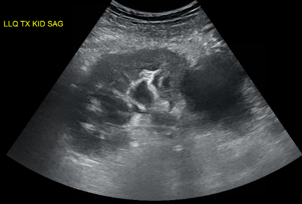
We were evaluating a kidney transplant recipient three weeks after surgery who presented with a rise in creatinine. We obtained an ultrasound which showed a large fluid collection and mild hydronephrosis. Prograf level was at goal. 
Peritransplant fluid collections may be produced by lymphoceles, urinomas, hematomas or abcesses. These collections may compress the ureter leading to hydronephrosis or may compress the renal vein causing a “compartment-like syndrome”. Since his creatinine was risen, we decided to drain the fluid collection. An important point is that mild hydronephrosis is reported in a large portion of kidney transplant ultrasounds and intervention is usually driven by the clinical setting. When in doubt, serial ultrasounds indicating progressive dilatation or the use of nuclear medicine imaging showing normal perfusion and parenchymal uptake but pooling of tracer in the renal pelvis with prolonged pelvic retention helps in confirming the diagnosis. 
His INR was 2.1 due to coumadin for a history of multiple clotted fistula. The interventional radiologist requested that we give FFP to reduce his INR to below 1.5 before considering the procedure. It was late Friday so we got 4U of FFP in and repeated INR. We were hoping that 4U of FFP would quickly reverse his mildly elevated INR. Interestingly, data supporting that is lacking. 
FFP can have an INR as high as 1.5 and transfusion of FFP will have little effect on minimally elevated INRs (Gearoid actually pointed that out in rounds). A study of the use of FFP in 121 patients with an INR in the range of 1.1 to 1.85, showed that correction of the INR to normal was achieved in only one patient (0.8 percent) and correction at least halfway to normal in only 15 percent. The median decrease in the PT and INR was 0.2 seconds and 0.07, respectively, and was independent of the number of units of FFP infused (median 2 units; range 1 to 20). Thus, available studies do not support the efficacy of FFP in treating bleeding or as prophylaxis for invasive procedures in patients with a mild coagulopathy (ie, INR below 2.0). 
After some discussion with IR, they agreed to go ahead with the procedure with an INR 1.9. About 600cc of yellow fluid was drained. Fluid creatinine was checked and was similar to serum level, ruling out an urinoma. This patient’s fluid collection was a lymphocele, which is the most common cause of peritransplant fluid collection due to disruption of renal lymphatics. It usually occurs weeks after surgery and drainage is required only in cases of suspected obstruction. With time, the amount of fluid collection tends to subside. Soon after drainage, his creatinine started to come down confirming the cause of his worsening renal function.

Figure: large peri-transplant fluid collection with associated hydronephrosis.

Friday, May 4, 2012

Still mysterious: the elusive circulating factor for FSGS


Important new findings were recently published in relation to proteinuria and FSGS, which are definitely of interest to our community.

First, the punch line:

There is new evidence for a “circulating factor” in recurrent FSGS in a fascinating case of a re-transplanted kidney (here)

BUT

There is growing evidence that suPAR is a non-specific marker of kidney disease and therefore not likely to be the “circulating factor.”(here)
In fact, it appears that it is non-specifically found in CKD, and correlates with a declining GFR.


Now for some details:

The re-transplanted kidney

A letter to the NEJM editor (here) describes an amazing case of resolution of recurrent FSGS after re-transplantation. 

A 27 year old patient with primary FSGS receiving a kidney from his healthy 24 year old sister developed proteinuria in the nephrotic range (up to 25 g/day!) within 2 days of transplantation, and had no improvement after plasmapheresis and standard immunosuppressive treatment. A renal biopsy confirmed foot process effacement, the first hallmark of recurrent podocyte damage heralding recurrent FSGS. Incredibly, with all appropriate consents and institutional approval, the transplant team removed the allograft from Patient 1 and re-transplanted it into another patient who had ESRD due to diabetes. Within 3-4 days, the proteinuria resolved and a repeat biopsy showed resolution of foot process effacement and re-establishment of a normal podocyte architecture. Eight months later, Patient 2 is reported to be doing very well, with good allograft function and no proteinuria.

This case demonstrates in a remarkable way that recurrent FSGS results from an elusive “factor” rapidly produced by the recipient (with primary FSGS), and that the allograft itself can remain fully functional if removed from the influence of this “factor” and placed in another patient.

suPAR is not suPER specific

What may have seemed to be exciting news in 2011, namely the notion that soluble uPAR may be predictive of recurrent FSGS (here), appears to be unfortunately evolving into yet another unsuccessful attempt to identify the ever elusive circulating factor.

Recent work published in Kidney International by Maas et al. (here) confirms that suPAR is not able to distinguish between idiopathic FSGS, secondary FSGS or minimal change disease. 

This is actually not surprising, because a closer look at the clinical data in Wei et al. (here) reveals that the admittedly arbitrary cut-off for separating primary FSGS from all other glomerular disease (3000 pg/ml) did not hold up when tested among their patient cohorts with idiopathic, recurrent versus non-recurrent FSGS (all had suPAR> 3000 pg/ml, thus suPAR could not predict the recurrent from the non-recurrent cases). 

The second figure in the Maas et al. paper may help explain this conundrum: they show a negative correlation between suPAR and eGFR, meaning that as GFR drops, suPAR levels rise, which essentially means that suPAR is simply a marker of CKD.

Future work will no doubt continue to address these issues, but the apparent lack of specificity of suPAR for FSGS casts serious doubt on its proposed role as the circulating factor.

So, the search is still on!!!

Thursday, May 3, 2012

Acute rejection: what do the circulating cells have to say about it?


One of the frequent situations that we face in the renal transplant clinic is the patient in otherwise good condition who presents with a slight rise in serum creatinine. Although this is sometimes due to reversible causes, such as high CNI levels or dehydration, acute rejection is of course in the differential. 
The fact that we still rely on an invasive procedure – the graft biopsy – for formal diagnosis of rejection clearly limits our ability for repetitive monitoring and potentially delays treatment. There is no doubt that a simple, non-invasive assay to monitor the immune status would be of great help in the day-to-day practice. Indeed this is currently a field of intense research in transplantation. We recently provided new insights into this issue. We optimized a simple assay to determine the level of activation of circulating blood mononuclear cells in renal transplant recipients. The method is relatively straightforward: peripheral blood is collected, cells are isolated and incubated overnight; cytokine production by the cultured cells is measured in the cell supernatant. The main objective was to determine if this assay, when used in patients for whom a biopsy was performed for an acute rise in serum creatinine, could identify those that would show histological signs of rejection. We found that the measurement of a single cytokine, IL-6, can predict rejection with a sensitivity of 92% and specificity of 63%. This tool could thus potentially be used to exclude rejection, which would be particularly helpful for low-risk or remote patients. 
Where do we go now? 
This work is a first step towards the development of a clinically useful tool. Ideally, a non-invasive test would be able to identify acute rejection well before the serum creatinine starts to rise. To achieve this, we now need to collect blood samples and study cell activation serially post transplant. What we need to determine more precisely is when the cells become activated before the usual signs of graft dysfunction occur. This will allow us to identify rejection early and by doing so, to prevent further graft damage. Although this sounds simple, from a research perspective this next step implies an enormous investment of human and lab resources. 
Sacha De Serres
Leonardo Riella 

Wednesday, May 2, 2012

To transplant or not to transplant: That is the question

A young woman presented for altruistic kidney donor evaluation. She had no significant past medical history other than two normal prior pregnancies. Physical examination was unremarkable. A CT angiogram revealed bilateral irregularities of the renal arteries and a 0.9 mm aneurysmal dilatation of right renal artery seen in the figure. Home blood pressures were normal and labs were otherwise unremarkable.

Clinical Question: In an asymptomatic, normotensive young adult female with an incidental finding of fibromuscular dysplasia (FMD) with bilateral renal artery involvement is it safe to proceed with altruistic kidney donation?

FMD is seen in approx 4% of patients worked up for kidney donation. The natural history is that in the majority of patients the lesions are non-progressive. In one study, primary and assisted primary patency rates were 66% and 87% at 5 years and the restenosis rate was 28% at 5 years. Hypertension improved or resolved within 3 months in 72% of patients and this was maintained in 73% at 5 years. After surgical revascularization long-term blood pressure control was maintained in 93% of patients and improvement or stabilization of renal function was noted in 92%.

Anecdotal case reports and case series suggest that despite patients being normotensive at the time of kidney donation patients with FMD can develop future radiological worsening of renal artery lesions, hypertension or ischemic renal disease. Kidney donors with FMD thus are potentially more likely to develop hypertension compared to kidney donors without FMD.

In the case discussed above, given the young age of the patient, bilateral nature of lesions and the presence of a renal aneurysm we recommended against altruistic kidney donation. We were also concerned that the recipient might develop hypertension post transplant given the irregularities that would be present in the transplanted renal artery.

The evidence would suggest that aneurysms that are >1.0 cm, non-calcified or found in pre-menopausal women are at high risk of rupture. Our patient had 2 out of these 3 criteria and therefore underwent coiling of the aneurysm with excellent results.

Author: Gautam Phadke, MD